. 2019 Mar 1;294(9):3294-3310.
doi: 10.1074/jbc.RA118.005685. Epub 2019 Jan 15.

Elucidation of the functional roles of the Q and I motifs in the human chromatin-remodeling enzyme BRG1

Affiliations

Elucidation of the functional roles of the Q and I motifs in the human chromatin-remodeling enzyme BRG1

Helen Hoffmeister et al. J Biol Chem. .

Abstract

The Snf2 proteins, comprising 53 different enzymes in humans, belong to the SF2 family. Many Snf2 enzymes possess chromatin-remodeling activity, requiring a functional ATPase domain consisting of conserved motifs named Q and I-VII. These motifs form two recA-like domains, creating an ATP-binding pocket. Little is known about the function of the conserved motifs in chromatin-remodeling enzymes. Here, we characterized the function of the Q and I (Walker I) motifs in hBRG1 (SMARCA4). The motifs are in close proximity to the bound ATP, suggesting a role in nucleotide binding and/or hydrolysis. Unexpectedly, when substituting the conserved residues Gln758 (Q motif) or Lys785 (I motif) of both motifs, all variants still bound ATP and exhibited basal ATPase activity similar to that of wildtype BRG1 (wtBRG1). However, all mutants lost the nucleosome-dependent stimulation of the ATPase domain. Their chromatin-remodeling rates were impaired accordingly, but nucleosome binding was retained and still comparable with that of wtBRG1. Interestingly, a cancer-relevant substitution, L754F (Q motif), displayed defects similar to the Gln758 variant(s), arguing for a comparable loss of function. Because we excluded a mutual interference of ATP and nucleosome binding, we postulate that both motifs stimulate the ATPase and chromatin-remodeling activities upon binding of BRG1 to nucleosomes, probably via allosteric mechanisms. Furthermore, mutations of both motifs similarly affect the enzymatic functionality of BRG1 in vitro and in living cells. Of note, in BRG1-deficient H1299 cells, exogenously expressed wtBRG1, but not BRG1 Q758A and BRG1 K785R, exhibited a tumor suppressor-like function.

Keywords: ATP; ATPase; ATPase domain; BRG1; SF2 helicase family; cell cycle; cell proliferation; chromatin remodeling; nucleosome; tumor suppressor gene.


Conflict of interest statement

The authors declare that they have no conflicts of interest with the contents of this article

Figures

Figure 1.
Figure 1.
Structure and gel analysis of hBRG1. A, sequence of the ATPase, i.e. residues 726–1249 from BRG1 isoform 2 (Uniprot-ID P51532-2). The ATP-binding domain (light blue) and the helicase C-terminal domain (dark blue) are highlighted, and conserved blocks (cons. bl.) and motifs (9) are indicated. The highly conserved Gln758 and Lys785 residues are printed in bold. B, homology model of the ATP-binding domain. The localization of ATPase motifs is indicated according to the color scheme shown on the right. ATPγS (black sticks) is docked into the ATP-binding site. C, a magnified view on the ATP-binding site, showing the orientation of the Q (violet) and I (red) motifs toward ATPγS. Gln758, Lys785, and ATPγS are shown as sticks, representing nitrogen (blue), sulfur (yellow), phosphorus (orange), oxygen (red), hydrogen (white), and carbon (green) atoms. Green dashed lines indicate hydrogen bonds between Gln758 and N6/7 of the nucleotide, and light blue lines indicate putative contacts between Lys785 and the phosphates of the nucleotide, which are presumably mediated by a Mg2+ atom (13, 10). The length of the respective bonds, including the hydrogen and the putative hydrogen acceptor, is listed. D, SDS-PAGE of BRG1 proteins. 1 μg of BRG1 proteins (wtBRG1, lanes 4 and 6; and mutants, lanes 1–3 and 7–10) were loaded on a 6% SDS-PAGE gel and stained with Coomassie (protein size marker, M, lane 5).
Figure 2.
Figure 2.
ATP binding and hydrolysis of wtBRG1 and mutants. A, ATP binding of WT BRG1 and mutants. 200 nm BRG1 proteins were incubated with 0.375 μCi of [γ-32P]ATP. The reactions were spotted on a nitrocellulose membrane. Bound [γ-32P]ATP was quantified after exposure on a phosphoimaging screen. Excess of nonradioactive ATP (1 and 100 μm) served as a competitor to verify signal specificity. Error bars represent the standard deviation of three technical replicates. B, ATPase activity of wtBRG1 and mutants. 250 nm of BRG1 proteins were incubated with 500 μm ATP in the presence of 0.1 μCi [γ-32P]ATP. ATP hydrolysis was stimulated with 400 nm centrally positioned mononucleosomes (77-NPS-77). Released (32PO4)3− was separated by TLC and quantified using phosphoimaging plates. Error bars represent the standard deviation of three technical replicates (−, absence of nucleosomes; +, presence of nucleosomes; background, chemical ATP background hydrolysis).
Figure 3.
Figure 3.
Nucleosome binding of WT BRG1 and mutants. 527 nm wtBRG1 or BRG1 mutants were incubated with streptavidin beads, with and without immobilized, biotinylated 77-NPS-77 nucleosomes (see also “Experimental procedures”). 20% of the IP reactions were used for gel analysis (lanes 9–16). The input samples (lanes 1–7) for gel analysis contained 0.21 μg of nucleosomes, 1% (final concentration) gelatin, 2 μg of BSA, or 0.96 μg of remodeler. All samples were loaded on 4–12% Bis-Tris PAA gels, whereby the upper part was stained with Coomassie and subsequently silver. The lower part of the gel was used for a Western blot with anti-H2B to verify the presence/release of nucleosomes. All IP reactions resolved in this figure originate from the same reaction.
Figure 4.
Figure 4.
Chromatin-remodeling activity of wtBRG1 and mutants and nucleotide binding to wtBRG1 in the presence and absence of nucleosomes-working model for the function of the Q and I motifs. A, 130 nm edge positioned mononucleosomes (upper panel, 0-NPS-77; lower panel, 77-NPS-77) were incubated with 250 or 500 nm enzyme with/without 1 mm ATP (left and right upper panel, lanes 2–4 and 14–16 with wtBRG1 and lanes 5–12 and 17–24 with indicated BRG1 mutants; lanes 1 and 13 with input nucleosome fraction and left and right lower panel, lanes 26–28 and 38–40 with wtBRG1 and lanes 29–36 and 41–48 with indicated BRG1 mutants; lanes 25 and 37 with input nucleosome fraction). Changes in nucleosome position were resolved on native 0.4× TBE, 6% PAA gels. The intensity of bands in each lane was quantified using Fuji Film multigauge software. Signal intensity profiles of selected gel lanes are highlighted with black triangles and numbers. The gel lane for the wtBRG1-containing reaction with ATP is displayed horizontally above the profiles, and triangles label the bands, corresponding to the peaks below. B, 200 nm wtBRG1 were incubated with 0.375 μCi of [γ-35S]ATP in the presence and absence of 400 nm or 600 nm nucleosomes. The reactions were spotted on a nitrocellulose membrane. Bound [γ-35S]ATP was quantified after exposure on a phosphoimaging screen. Equivalent amounts of rabbit IgG and nucleosomes served as controls. Error bars represent the standard deviation of three technical replicates. C, mutating the conserved amino acids of the Q and I motifs did not alter the ability of the enzyme to bind ATP, and also the basal ATPase activity was retained to a level comparable with wtBRG1. All mutants were still capable to interact with nucleosomes like wtBRG1. However, their nucleosome-stimulated ATPase activity was lost. Accordingly, the chromatin-remodeling activity of all mutants was impaired, with the exception of edge-positioned nucleosomes, in which the basal ATP hydrolysis rates of the BRG1 mutants (see also Fig. 2B) were sufficient to result in a “slight” nucleosome repositioning (see also Fig. 4A). However, an efficient nucleosome remodeling only took place in the reactions containing wtBRG1, the sole protein, being significantly stimulated in its ATP hydrolysis rate upon nucleosome addition (Fig. 2B). This suggests that either the current speed and/or the final amount of the released energy in the course of high ATP hydrolysis rates are required for “efficient” nucleosome translocation (initial, edge-positioned nucleosomes are presented in dark gray; weakly (left) and efficient (right) remodeled nucleosomes are displayed in distinct, lighter shades of gray). Because we can exclude an influence of ATP or nucleosomes on the binding of the respective other substrate to BRG1, we therefore specify the function of both motifs as follows: the Q and I motifs are transmitting the stimulatory impulse of the (remodeler) associated nucleosome to the ATP hydrolysis center of the enzyme, stimulating in turn the chromatin-remodeling rate of the protein.
Figure 5.
Figure 5.
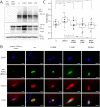
Western blotting, immunocytochemical, and cell cycle analysis of H1299 cells expressing wtBRG1 and mutant proteins. A, 48 h after transient transfection with the indicated plasmids, 40 μg of whole cell extracts of H1299 cells were loaded on 6.5% SDS gels, which were subsequently transferred to PVDF membranes via semidry blotting. The PVDF membrane was cut into three pieces, which were incubated with the indicated antibodies. Tubulin served as a loading control. B, 48 h after transient transfection with the indicated plasmids, H1299 cells were fixed in 4% PFA, Triton X-100–permeabilized, and immunostained with an “anti-FLAG” primary antibody in combination with an Alexa 488–labeled secondary antibody. The nuclear DNA was colored via DAPI. The cells were finally mounted in PBS/glycerol, and pictures for the indicated fluorophores were taken at the fluorescence microscope Axiovert 200M. Scale bar, 20 μm. C, 24 h after transient transfection with the indicated plasmids, H1299 cells were seeded (subconfluent) for a period of 6 days on P150 plates. Six days after transfection, the cells were labeled with Hoechst, trypsinized, filtered, and finally analyzed with a CyFLOW space flow cytometer (Partec). The ratio between the cell numbers between the G1 and G2 populations (see “Experimental procedures”) from five or six biological replicates was calculated (G1/G2 ratio) and plotted as box-plot diagrams, using the software Origin 2017 (vc, vector control = empty mCherry vector). The median is shown as a line, and the average is shown as a green box and is displayed right to the box. The statistical significance was calculated in Origin 2017 software, performing a t test with two samples, mean 1 − mean 2 <> 0, significance-niveau 0.05, assuming heteroscedastic variance (Welch Korrektur) with *, p < 0.05; **, p < 0.01; and ***, p < 0.001 and not significant (n.s.).

References

    1. Luger K., Mäder A. W., Richmond R. K., Sargent D. F., and Richmond T. J. (1997) Crystal structure of the nucleosome core particle at 2.8 A resolution. Nature 389, 251–260 10.1038/38444 - DOI - PubMed
    1. Clapier C. R., and Cairns B. R. (2009) The biology of chromatin remodeling complexes. Annu. Rev. Biochem. 78, 273–304 10.1146/annurev.biochem.77.062706.153223 - DOI - PubMed
    1. Huang R. C., and Huang P. C. (1969) Effect of protein-bound RNA associated with chick embryo chromatin on template specificity of the chromatin. J. Mol. Biol. 39, 365–378 10.1016/0022-2836(69)90323-4 - DOI - PubMed
    1. Porter I. M., Khoudoli G. A., and Swedlow J. R. (2004) Chromosome condensation: DNA compaction in real time. Curr. Biol. 14, R554–R556 10.1016/j.cub.2004.07.009 - DOI - PubMed
    1. Rodríguez-Campos A., and Azorín F. (2007) RNA is an integral component of chromatin that contributes to its structural organization. PLoS One 2, e1182 10.1371/journal.pone.0001182 - DOI - PMC - PubMed

Publication types

Associated data

LinkOut - more resources