. 2019 May;13(5):1239-1251.
doi: 10.1038/s41396-019-0344-9. Epub 2019 Jan 15.

Bacterial persistence promotes the evolution of antibiotic resistance by increasing survival and mutation rates

Affiliations

Bacterial persistence promotes the evolution of antibiotic resistance by increasing survival and mutation rates

Etthel Martha Windels et al. ISME J. 2019 May.

Abstract

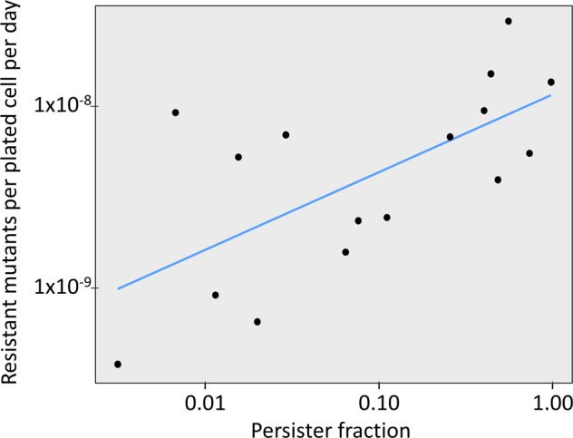
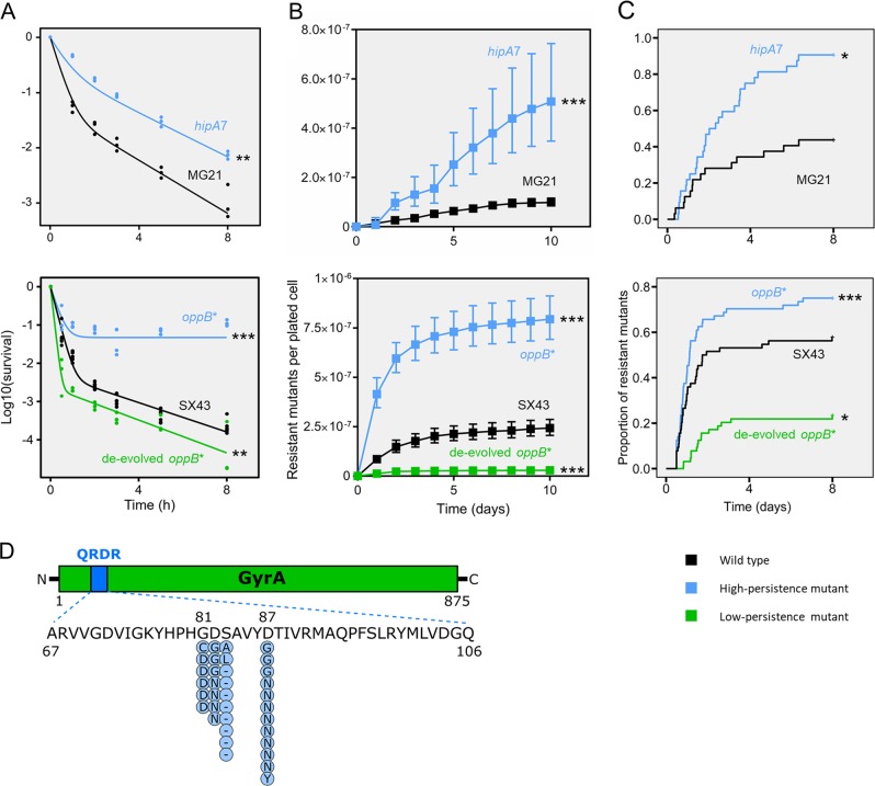
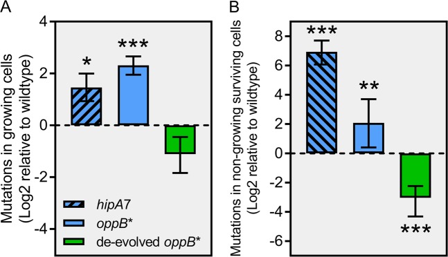
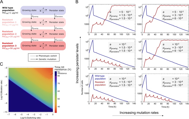
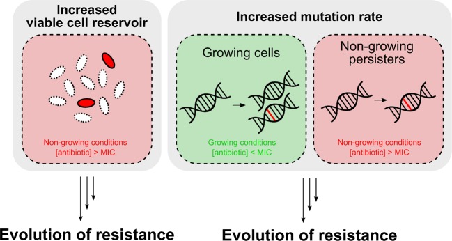
Persisters are transiently antibiotic-tolerant cells that complicate the treatment of bacterial infections. Both theory and experiments have suggested that persisters facilitate genetic resistance by constituting an evolutionary reservoir of viable cells. Here, we provide evidence for a strong positive correlation between persistence and the likelihood to become genetically resistant in natural and lab strains of E. coli. This correlation can be partly attributed to the increased availability of viable cells associated with persistence. However, our data additionally show that persistence is pleiotropically linked with mutation rates. Our theoretical model further demonstrates that increased survival and mutation rates jointly affect the likelihood of evolving clinical resistance. Overall, these results suggest that the battle against antibiotic resistance will benefit from incorporating anti-persister therapies.


Conflict of interest statement

The authors declare that they have no conflict of interest.

Figures

Fig. 1
Fig. 1
Across natural E. coli isolates, persister fractions are strongly predictive of the likelihood to evolve genetic antibiotic resistance. Persister fractions (n = 3 per strain) and the average number of resistant mutants per plated cell per day (n = 5 per strain) on ciprofloxacin-containing Mueller-Hinton agar (4x MIC) are plotted for 16 ECOR isolates on a log-log scale. To account for the phylogenetic non-independence among strains, PGLS regression was used to fit a linear model onto the log-log transformed data (R² = 0.498, p = 0.002)
Fig. 2
Fig. 2
Ciprofloxacin resistance development is increased in high-persistence mutants and decreased in a low-persistence mutant. a Ciprofloxacin time-kill curves of stationary phase cultures show increased persister fractions in hipA7 and oppB*, and decreased persistence in de-evolved oppB*. Individual data points from three independent replicates are shown. Lines represent a biphasic exponential fit to the data. Parameter estimates for persister fractions were compared with Wald F-tests. b Cumulative number of resistant mutants per plated cell on Mueller-Hinton agar supplemented with ciprofloxacin (2x MIC). Data points represent averages ± s.e.m. (n = 10). Statistical comparison was done by two-sided t-tests on coefficients of a log-linear model. c Cumulative proportion of wells with ciprofloxacin-containing Mueller-Hinton broth (2x MIC) in which resistant mutants emerged (n = 32-64). Statistical comparison was done by Wald tests on coefficients of a Cox proportional hazards model, to take into account censoring. d Amino acid changes caused by gyrA mutations detected in ciprofloxacin-resistant colonies. *p ≤ 0.05; **p ≤ 0.01; ***p ≤ 0.001, and ns non-significant
Fig. 3
Fig. 3
Persistence is correlated with elevated mutation rates. a Mutation rate (mutations per cell per division) of growing cells determined from the number of ciprofloxacin-resistant mutants after two days using fluctuation analysis, relative to the wild type. Error bars show 95% c.i. (n = 10), p-values are from one-sided t-tests. b Mutations in non-growing cells surviving lethal antibiotic exposure (mutations per cell per day), relative to the wild type. Error bars show 95% c.i.’s (n = 10), p-values are from one-sided t-tests. *p ≤ 0.1; **p ≤ 0.05; ***p ≤ 0.01
Fig. 4
Fig. 4
A mathematical simulation model shows that increased persistence and mutation rates jointly drive the evolution of resistance under clinical conditions. a Schematic diagram of the mathematical model showing all possible transitions between the different genotypes and phenotypic states. b Population dynamics for wild type and resistant cells during antibiotic treatment for increasing persister fractions with or without associated increased mutation rates. The separate dynamics of all different subpopulations under the same sets of parameters are given in Fig. S10. c Heat map of the time at which the resistant cells take over the population (>108 resistant cells), as a function of the mutation rate and the persister fraction (switching rate). The mutation rate is shown for normal cells, the mutation rate for persisters is 104-fold higher. See text and Supplementary Methods for details on the mathematical model
Fig. 5
Fig. 5
Proposed model explaining how persistence accelerates the evolution of genetic resistance. Persistence refers to the fraction of cells in a sensitive population that survives otherwise lethal antibiotic exposure. High persistence accelerates the evolution of antibiotic resistance by increasing the number of viable cells available for mutation, thus increasing the likelihood for a resistance-conferring mutation to arise. Additionally, persistence is linked with higher mutation rates in growing cells and cells that survive lethal antibiotic exposure but cannot grow (persisters). Together, these two factors have the potential to accelerate the evolution of antibiotic resistance in high-persistence strains in different environments with or without antibiotics. Red depicts environments where the antibiotic concentration is above the MIC, while green depicts environments with subinhibitory or zero antibiotic concentrations

References

    1. Davies J, Davies D. Origins and evolution of antibiotic resistance. Microbiol Mol Biol Rev. 2010;74:417–33. doi: 10.1128/MMBR.00016-10. - DOI - PMC - PubMed
    1. World Health Organization. Antimicrobial resistance: global report on surveillance. WHO press, Geneva, Switzerland; 2014.
    1. Brauner A, Fridman O, Gefen O, Balaban NQ. Distinguishing between resistance, tolerance and persistence to antibiotic treatment. Nat Rev Microbiol. 2016;14:320–30. doi: 10.1038/nrmicro.2016.34. - DOI - PubMed
    1. Bumann D. Heterogeneous host-pathogen encounters: act locally, think globally. Cell Host Microbe. 2015;17:13–19. doi: 10.1016/j.chom.2014.12.006. - DOI - PubMed
    1. Conlon BP. Staphylococcus aureus chronic and relapsing infections: evidence of a role for persister cells. Bioessays. 2014;36:991–6. doi: 10.1002/bies.201400080. - DOI - PubMed

Publication types