. 2018 Dec 16:2018:3523642.
doi: 10.1155/2018/3523642. eCollection 2018.

Involvement of M1 Macrophage Polarization in Endosomal Toll-Like Receptors Activated Psoriatic Inflammation

Affiliations

Involvement of M1 Macrophage Polarization in Endosomal Toll-Like Receptors Activated Psoriatic Inflammation

Chih-Hao Lu et al. Mediators Inflamm. .

Abstract

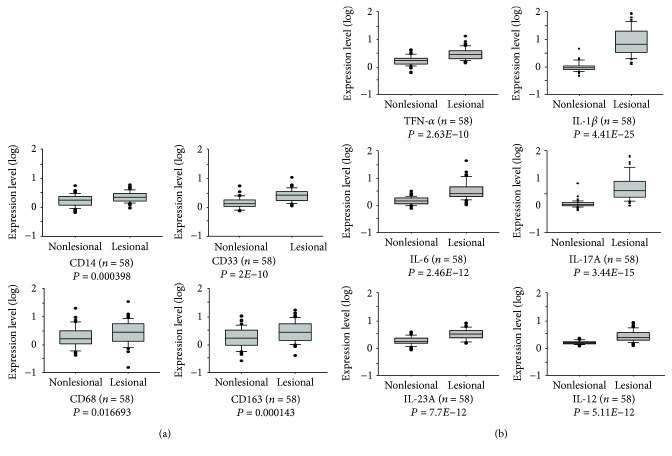
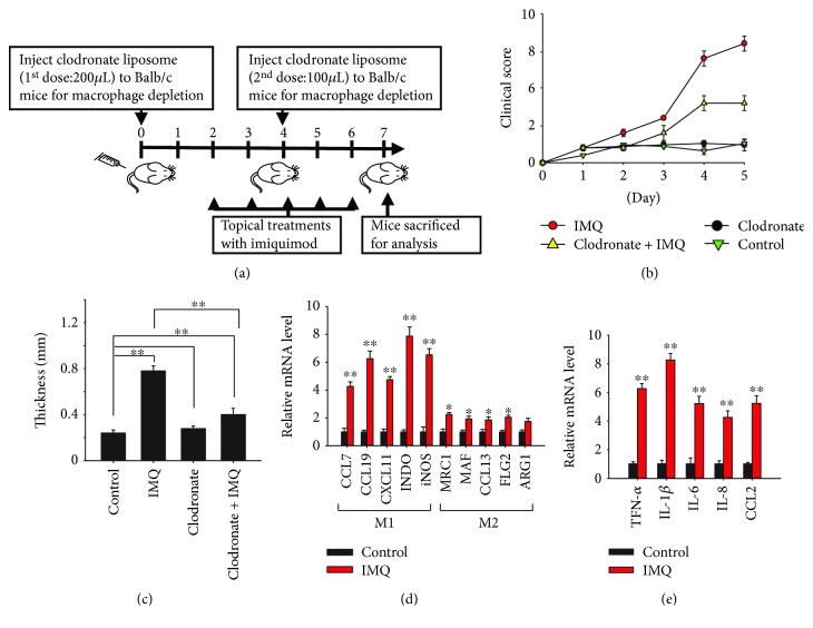
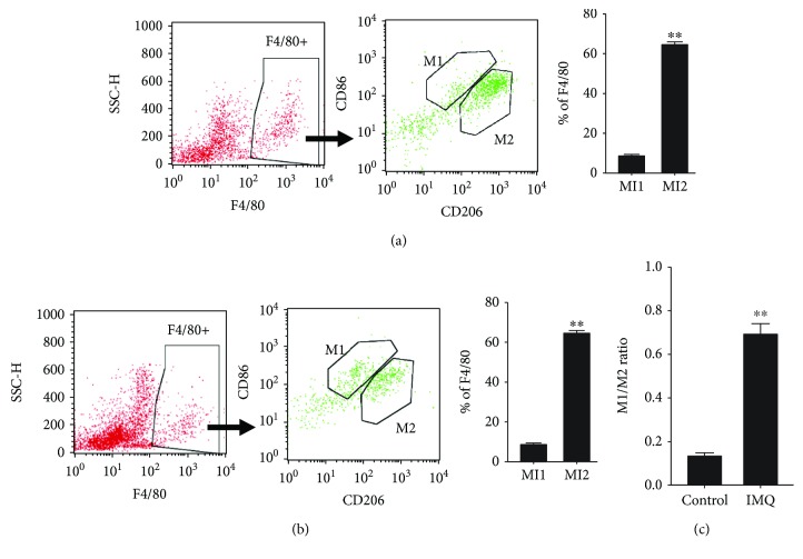
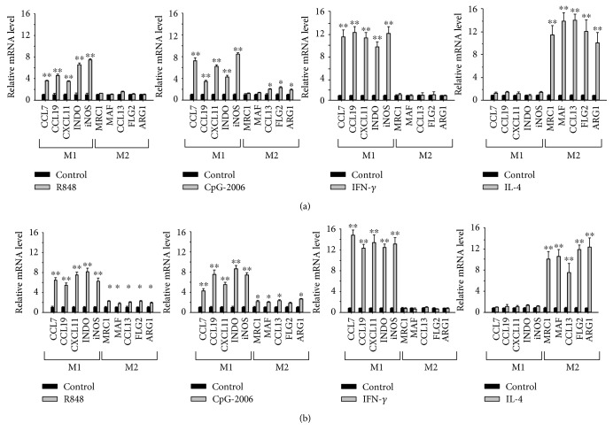
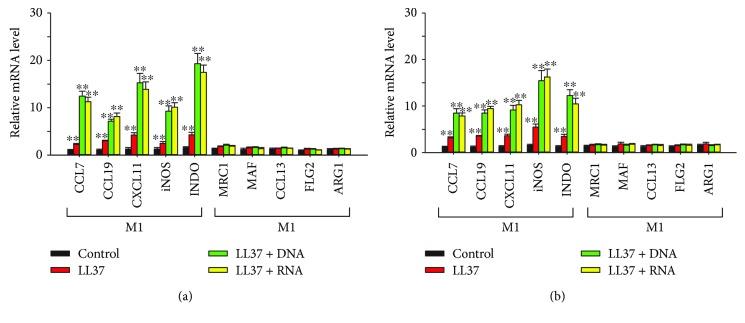
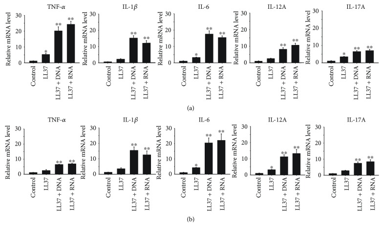
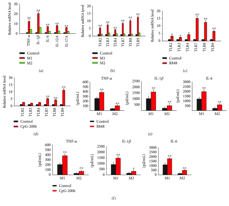
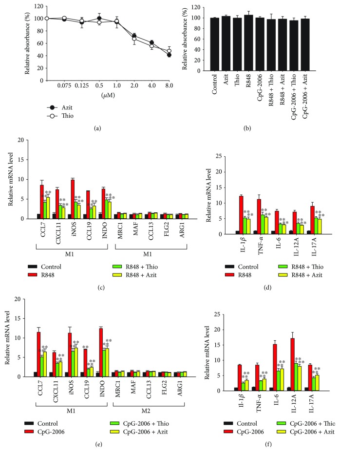
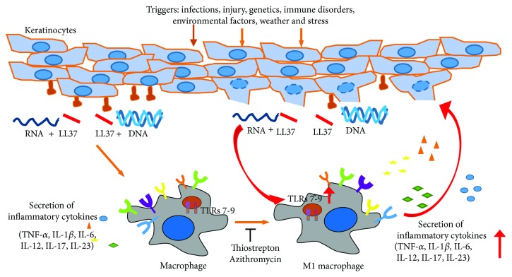
Psoriasis is a chronic inflammatory skin disorder that affects ~2%-3% of the worldwide population. Inappropriate and excessive activation of endosomal Toll-like receptors 7, 8, and 9 (TLRs 7-9) at the psoriatic site has been shown to play a pathogenic role in the onset of psoriasis. Macrophage is a major inflammatory cell type that can be differentiated into phenotypes M1 and M2. M1 macrophages produce proinflammatory cytokines, and M2 macrophages produce anti-inflammatory cytokines. The balance between these two types of macrophages determines the progression of various inflammatory diseases; however, whether macrophage polarization plays a role in psoriatic inflammation activated by endosomal TLRs has not been investigated. In this study, we investigated the function and mechanism of macrophages related to the pathogenic role of TLRs 7-9 in the progression of psoriasis. Analysis of clinical data in database revealed significantly increased expression of macrophage markers and inflammatory cytokines in psoriatic tissues over those in normal tissues. In animal studies, depletion of macrophages in mice ameliorated imiquimod, a TLR 7 agonist-induced psoriatic response. Imiquimod induced expression of genes and cytokines that are signature of M1 macrophage in the psoriatic lesions. In addition, treatment with this TLR 7 agonist shifted macrophages in the psoriatic lesions to a higher M1/M2 ratio. Both of the exogenous and endogenous TLR 7-9 ligands activated M1 macrophage polarization. M1 macrophages expressed higher levels of proinflammatory cytokines and TLRs 7-9 than M2 macrophages. These results suggest that by rendering macrophages into a more inflammatory status and capable of response to their ligands in the psoriatic sites, TLR 7-9 activation drives them to participate in endosomal TLR-activated psoriatic inflammation, resulting in an amplified inflammatory response. Our results also suggest that blocking M1 macrophage polarization could be a strategy which enables inhibition of psoriatic inflammation activated by these TLRs.


Figures

Figure 1
Figure 1
Elevated expression of monocytes and macrophage markers and inflammatory cytokines in human psoriatic lesions. Gene expression data in Gene Expression Omnibus (GEO) dataset GSE13355 were analyzed for (a) expression of monocyte and/or macrophage markers and (b) inflammatory cytokines in tissue with and without lesions from psoriatic patients (n = 58).
Figure 2
Figure 2
Depletion of macrophage attenuates imiquimod- (IMQ-) induced psoriasis-like inflammation. (a) Balb/c mice were intraperitoneally injected with clodronate-containing liposomes and topically treated with IMQ cream following the schedule illustrated. (b) The severity of inflammatory responses on the skin was assessed on the basis of the Psoriasis Area Severity Index. (c) Five days after IMQ treatment, the mice were sacrificed and their skin thickness was measured with vernier caliper to assess the severity of the psoriatic responses. (d, e) Tissue samples from the control and IMQ-treated mice were analyzed using quantitative real-time polymerase chain reaction for the expression of (d) M1 and M2 macrophage markers and (e) inflammatory cytokines and chemokines. The data represent mean ± standard deviation (n = 5); P < 0.05 and ∗∗ P < 0.01 compared with the controls, or as indicated.
Figure 3
Figure 3
Increased M1/M2 macrophage ratio in the imiquimod-induced psoriatic lesions. Balb/c mice were treated with imiquimod (IMQ) for 5 days as illustrated in Figure 2(a). Macrophages in the tissues were analyzed by flow cytometer. (a, b) A set of representative histograms is shown for the gated population of F4/80-positive macrophages, F4/80 CD86 double-positive M1 macrophages, and F4/80 CD206 double-positive M2 macrophages in tissues from (a) control and (b) IMQ-treated mice. (c) The ratio of M1 and M2 macrophages in tissues from control- and IMO-treated mice was calculated from the bar figures in (a) and (b). Bar figures: the data represent mean ± standard deviation (n = 5), ∗∗ P < 0.01.
Figure 4
Figure 4
Induction of M1 macrophage polarization by endosomal Toll-like receptor (TLR) 7–9 ligands. (a) THP-1 macrophages and (b) bone marrow-derived macrophages were treated with 2 μM R848 or CpG-2006 for 24 h. In addition, these cells were treated with 20 ng/mL interferon- (IFN-) γ and interleukin- (IL-) 4 for 24 h for control of M1 and M2 macrophage polarization, respectively. Polarization of the macrophages into M1 and M2 phenotypes was determined by quantitative real-time polymerase chain reaction analysis for expression of their signature genes. Data represent mean ± standard deviation of three independent experiments; P < 0.05 and ∗∗ P < 0.01 compared with the controls.
Figure 5
Figure 5
Induction of M1 macrophage polarization by endogenous Toll-like receptor (TLR) 7–9 ligands. (a) THP-1 macrophages and (b) bone marrow-derived macrophages were treated with 2 μg/mL LL37, LL37/DNA, or LL37/RNA complex for 24 h. Polarization of the macrophages into M1 and M2 phenotypes was determined by quantitative real-time polymerase chain reaction (RT-qPCR) analysis for expression of their signature genes. Data represent mean ± standard deviation of three independent experiments; ∗∗ P < 0.01 compared with the controls.
Figure 6
Figure 6
Induction of cytokine production by endogenous Toll-like receptor (TLR) 7–9 ligands. (a) THP-1 macrophages and (b) bone marrow-derived macrophages were treated with 2 μg/mL LL37, LL37/DNA, or LL37/RNA complex for 24 h. Production of cytokines was determined by quantitative real-time polymerase chain reaction (RT-qPCR) analysis. Data represent mean ± standard deviation of three independent experiments; P < 0.05 and ∗∗ P < 0.01 compared with the controls.
Figure 7
Figure 7
High expression of proinflammatory cytokines and Toll-like receptors (TLRs) in M1 macrophages and induction of TLR 7–9 expression by their agonists in macrophages. THP-1 macrophages were treated with 20 ng/mL interferon- (IFN-) γ and interleukin- (IL-) 4 to polarize them into M1 and M2 macrophages. (a) Expression of genes for proinflammatory cytokines and (b) expression of TLRs in these cells were analyzed using quantitative real-time polymerase chain reaction (RT-qPCR). (c, d) To assess the capability of inducing the expression of TLRs 7–9 by their agonists, THP-1 macrophages were treated with 2 μM (c) R848 or (d) CpG-2006 for 24 h and expression of different TLRs was analyzed using RT-qPCR. (e, f) The IFN-γ- and IL-4-polarized M1 and M2 macrophages were treated with 2 μM (e) R848 or (f) CpG-2006 for 24 h, and production of cytokines as indicated in medium was measured with enzyme-linked immunosorbent assay. Data represent mean ± standard deviation of three independent experiments; P < 0.05 and ∗∗ P < 0.01 compared with the controls (c–f) or between M1 and M2 macrophages (a, b).
Figure 8
Figure 8
Thiostrepton and azithromycin attenuate Toll-like receptor (TLR) 7- to 9-induced M1 macrophage polarization and cytokine production in vitro and in vivo. (a, b) Bone marrow-derived macrophages (BMDMs) were treated with different concentrations of (a) thiostrepton (Thio) and azithromycin (Azit) and (b) 1 μM Thio or Azit with or without 2 μM R848 or CpG-2006 to assess the cytotoxicity of these treatments. (c–f) The cells were treated with 1 μM Thio or Azit plus 2 μM (c, d) R848 or (e, f) CpG-2006 for 24 h. Expression of signature genes for (c, e) M1 and M2 macrophages and expression of genes for (d, f) inflammatory cytokines were analyzed with quantitative real-time polymerase chain reaction (RT-qPCR). Data represent mean ± standard deviation of three independent experiments; ∗∗ P < 0.01 compared with the R848- or CpG-2006-treated group (c–f).
Figure 9
Figure 9
Model for the role of macrophages in the pathogenic role of Toll-like receptor (TLR) 7- to 9-activated psoriatic inflammation. Ligands of TLRs 7–9 activate cytokine production, TLR 7–9 expression, and M1 polarization in macrophages. M1 macrophages express higher levels of proinflammatory cytokines and TLRs 7–9. These render macrophages to be more inflammatory and further respond to the TLR ligands and lead to an amplification of TLR 7- to 9-activated inflammation at the psoriatic sites. Inhibitors of TLRs 7–9 such as thiostrepton and azithromycin block this TLR-activated M1 macrophage polarization, which can be a mechanism for their inhibitory activity in reducing psoriatic inflammation. Red arrows show the increased expression of TLRs 7–9 and proinflammatory cytokines.

Similar articles

Cited by

References

    1. Christophers E. Psoriasis--epidemiology and clinical spectrum. Clinical and Experimental Dermatology. 2001;26(4):314–320. doi: 10.1046/j.1365-2230.2001.00832.x. - DOI - PubMed
    1. Perera G. K., Di Meglio P., Nestle F. O. Psoriasis. Annual Review of Pathology. 2012;7(1):385–422. doi: 10.1146/annurev-pathol-011811-132448. - DOI - PubMed
    1. Lowes M. A., Suarez-Farinas M., Krueger J. G. Immunology of psoriasis. Annual Review of Immunology. 2014;32(1):227–255. doi: 10.1146/annurev-immunol-032713-120225. - DOI - PMC - PubMed
    1. Lowes M. A., Bowcock A. M., Krueger J. G. Pathogenesis and therapy of psoriasis. Nature. 2007;445(7130):866–873. doi: 10.1038/nature05663. - DOI - PubMed
    1. Chuang T. H., Ulevitch R. J. Identification of hTLR10: a novel human Toll-like receptor preferentially expressed in immune cells. Biochimica et Biophysica Acta (BBA) - Gene Structure and Expression. 2001;1518(1-2):157–161. doi: 10.1016/S0167-4781(00)00289-X. - DOI - PubMed

MeSH terms