. 2016 Jun;14(1):211-217.
doi: 10.1016/j.jgeb.2015.11.002. Epub 2015 Nov 25.

Inhibitory potential of important phytochemicals from Pergularia daemia (Forsk.) chiov., on snake venom (Naja naja)

Affiliations

Inhibitory potential of important phytochemicals from Pergularia daemia (Forsk.) chiov., on snake venom (Naja naja)

S T V Raghavamma et al. J Genet Eng Biotechnol. 2016 Jun.

Abstract

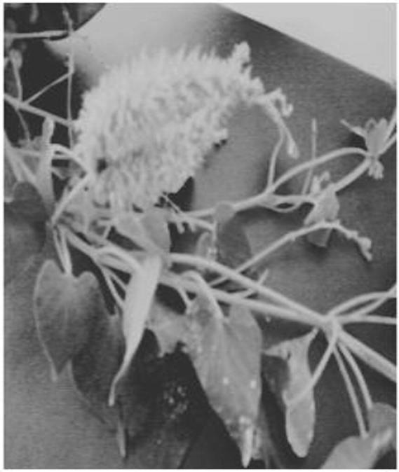
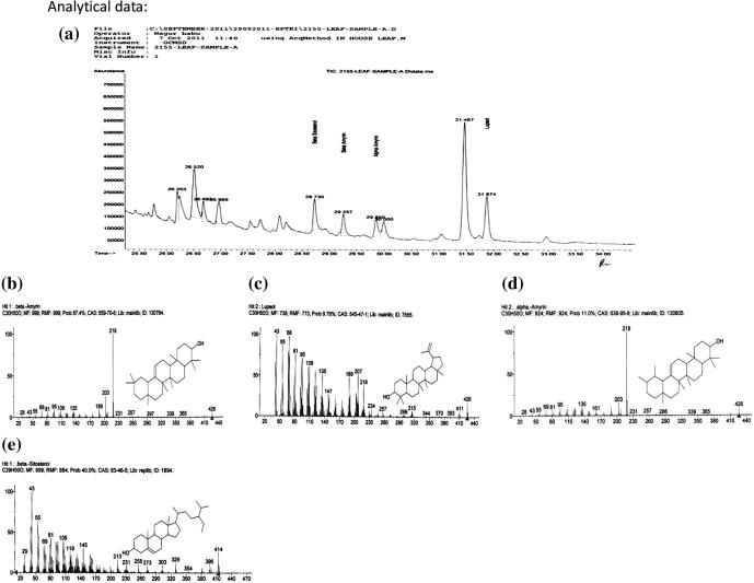
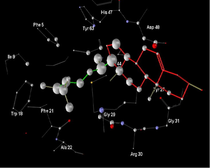
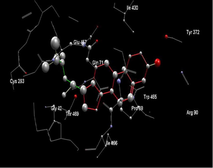
Pergularia daemia (Forsk.) chiov., is a milk weed of Asclepiadaceae family. In the present study β-sitosterol, β-amyrin, α-amyrin and lupeol were identified in the leaf by GC-MS. Molecular docking studies were performed to evaluate their activities on phospholipase A2 (PLA2) and l-amino acid oxidase enzymes which constituted a rich source in snake venoms (Naja naja). Snake venom Phospholipase A2 with PDB code 1A3D devoid of co-crystallized ligand was extracted from Protein Data Bank. Using Molegro Virtual Docker two cavities are formed by cocrystallization. l-Amino acid oxidase (PDB code 4E0V) was a receptor model with a co-crystallized ligand FAD. Among the phytochemicals analysed, β-sitosterol displayed high affinity of binding to the active site regions of phospholipase A2 and l-amino acid oxidase, respectively. The affinity of binding was -125.939 and -157.521 kcal/mole identified by gold scores. α-Amyrin and β-amyrin had two hydrogen bond interactions with PLA2. Hence this study suggests that β-sitosterol identified in P. daemia can antagonize PLA2 and LAAO activities and forms a theoretical basis for the folk use of the plant against snake venom.

Keywords: Molecular docking; Pergularia daemia; Phospholipase A2; Snake venom; l-Amino acid oxidase.


Figures

Figure 1
Figure 1
Twig of Pergularia daemia bearing flowers and fruit. The leaves are almost glabrous above and velvety below. The flower corolla forms a greenish-yellow or dull white tube. The fruits release ovate seeds covered with velvety hairs.
Figure 2
Figure 2
By using the Pergularia daemia trnL-trnF intergenic spacer, partial sequence; plastid a phylogenetic tree was constructed. Maximum likelihood phylogram was discovered in 8 independent ratchet analyses. Note the broken branch to the outgroup, Cynanchum, with the exception of a few weakly supported nodes, this topology is identical to the strict consensus of most parsimonious trees discovered.
Figure 4
Figure 4
GC–MS chromatogram of P. daemia leaf extract showing β-sitosterol, α-amyrin, β-amyrin and lupeol Chromatogram b, c, d, e shows compounds identified compared to inbuilt NIST library.
Figure 3
Figure 3
In-vitro PLA2 activity of the extract in agarose gel impregnated with 1.2% sheep erythrocytes and 1.2% egg yolk and 10 mM CaCl2. 1–8 Cavities show inhibition of haemolytic activity of naja venom by increasing dose of plant extract (5, 10, 15, 20, 25, 30, 35, 40 μl of plant extract) in series.
Figure 5
Figure 5
Tannic acid standard graph for the estimation of phenols from Pergularia daemia extract using the Folins method.
Figure 6
Figure 6
Standard graph of quercetin for the estimation of flavonoids from Pergularia daemia extract.
Figure 7
Figure 7
Graph shows percentage inhibition of PLA2 activity corresponding to phenolic content of the extract.
Figure 8
Figure 8
Venom protease inhibition activity of Pergularia daemia plotted against the corresponding concentration of the phenolic component in the extract.
Figure 9
Figure 9
Percentage inhibition of hyaluronidase activity of Pergularia daemia plotted against corresponding concentration of phenolic component in the extract.
Figure 10
Figure 10
Energy contributions of active site amino acids of 1A3D with β-sitosterol. Atoms contributing energy are represented as spheres (size corresponds to the energy associated).
Figure 11
Figure 11
Energy contributions of active site amino acids of 4E0V with β-sitosterol. Atoms contributing energy are represented as spheres (size corresponds to the energy associated).

Similar articles

Cited by

References

    1. Elango V., Ambujavalli L., Amala Basker E., Sulochana N. Fitoterapia. 1985;56:300–302.
    1. Singh V.P., Sharma S.K., Khare V.S. Indian Drugs Pharm. Ind. 1980;5:7–12.
    1. Dutta A., Ghosh S. J. Pharm. Sci. 1947;36:250–252. - PubMed
    1. Arseculeratne S.N., Gunatilaka A.A.L., Panabokke R.G. J. Ethnopharmacol. 1985;13(3) 323-3. - PubMed
    1. Selvanayahgam Z.E., Gnanevendhan S.G., Balakrishna K., Rao R.B. J. Herbs Spices Med. Plants. 1994;2:45–100.

LinkOut - more resources