Review
. 2018 Dec 28:7:F1000 Faculty Rev-1983.
doi: 10.12688/f1000research.16629.1. eCollection 2018.

Recent insights into the structure and function of Mitofusins in mitochondrial fusion

Affiliations
Review

Recent insights into the structure and function of Mitofusins in mitochondrial fusion

Mickael M Cohen et al. F1000Res. .

Abstract

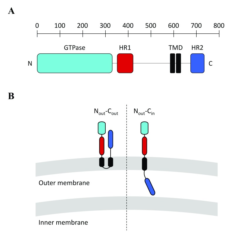
Mitochondria undergo frequent fusion and fission events to adapt their morphology to cellular needs. Homotypic docking and fusion of outer mitochondrial membranes are controlled by Mitofusins, a set of large membrane-anchored GTPase proteins belonging to the dynamin superfamily. Mitofusins include, in addition to their GTPase and transmembrane domains, two heptad repeat domains, HR1 and HR2. All four regions are crucial for Mitofusin function, but their precise contribution to mitochondrial docking and fusion events has remained elusive until very recently. In this commentary, we first give an overview of the established strategies employed by various protein machineries distinct from Mitofusins to mediate membrane fusion. We then present recent structure-function data on Mitofusins that provide important novel insights into their mode of action in mitochondrial fusion.

Keywords: Amphipathic Helix; Atlastin; Coiled-coil; Fusion; GTPase; Hemagglutinin; Heptad Repeat; Lipids; Membrane; Mitochondria; Mitofusin; SNARE.


Conflict of interest statement

No competing interests were disclosed.No competing interests were disclosed.No competing interests were disclosed.

Figures

Figure 1.
Figure 1.
( A) Molecular architecture of Mitofusin 1 (MFN1). Like MFN1, all Mitofusins include an N-terminal GTPase domain (light-blue) and two C-terminal heptad repeat domains, HR1 (red) and HR2 (dark blue), that sandwich a transmembrane region (black). The yeast Mitofusin Fzo1 includes an additional heptad repeat domain (HRN) located upstream of the GTPase domain (not depicted here). ( B) Possible topologies of Mitofusins. (Left) A transmembrane region with two transmembrane domains (TMDs) gives Mitofusins a topology in which the N- and C-terminal extremities are exposed to the cytoplasm (N out–C out topology). (Right) It was recently demonstrated that Mitofusins from vertebrates could also include a single TMD, which keeps the N-terminal GTPase and HR1 domains in the cytoplasm but places the C-terminal HR2 domain in the mitochondrial intermembrane space (N out–C in topology). Note that the BDLP1-like folding of Mitofusins observed in the X-ray structures of MFN1 ( Figure 2) is compatible with the N out–C out but not the N out–C in topology.
Figure 2.
Figure 2.. X-ray structure of a Mitofusin 1 (MFN1) fragment.
The fragment is composed of the predicted GTPase domain (purple and green) and the first ~15 N-terminal residues of the heptad repeat domain HR1 (red) linked to the last ~45 C-terminal residues of the HR2 domain (blue) via an artificial linker. The structure of this fragment, named mini-MFN1, consists of a typical G-domain (purple) and a four-helix bundle domain (HB1), which includes two helices from an N-terminal extension of the GTPase (green), the short N-terminal fragment of HR1 (red), and the C-terminal fragment of HR2 (blue). (Left) Structure of the “open-HB1” dimeric form of mini-MFN1 (Protein Data Bank entry 5GOM ) obtained upon addition of GDP/AlF 4 (but with only GDP in the crystal). (Right) Structure of the “closed-HB1” dimeric form of mini-MFN1 (Protein Data Bank entry 5YEW ) observed in the presence of GDP/BeF 3 . The indicated distances were measured between the N-terminal sides of HR2. The figures were prepared using Chimera.
Figure 3.
Figure 3.. Hypothetical mode of action of Mitofusin in mitochondrial fusion.
( A) Based on the available X-ray structures of Mitofusin 1 (MFN1) ( Figure 2) , , , as well as structural modeling of MFN1 and Fzo1 using BDLP1 as a template , , full-length Mitofusins should be constituted of four distinct structural motifs in their three-dimensional conformation: a G-domain (pink) followed by two sequential four-helix bundles, HB1 (green) and HB2 (yellow), and a transmembrane region that spans the outer membrane twice. ( B) Mitofusin molecules may dimerize across outer mitochondrial membranes upon GTP binding, which leads to long-distance (~20 nm) docking of mitochondria. GTP hydrolysis may then induce a large conformational rearrangement of Mitofusin, through either a “scissor-like” (top panel) or a “self-folding” (bottom panel) mechanism, which brings outer mitochondrial membranes in closer proximity (short-distance docking). These two docking states may be further stabilized by the formation of a ring of trans-Mitofusin complexes (not depicted here) at the periphery of the contact zone between mitochondria. Short-distance docking may also be reinforced by the formation of antiparallel trans-HR2 dimers (not shown for clarity). Mitochondrial fusion may proceed as a result of local membrane deformation near the TMD when Mitofusin undergoes its GTP hydrolysis-dependent conformational transition and membrane structure perturbation by the HR1 domain (symbolized by the red star).

References

    1. Gorsich SW, Shaw JM: Importance of mitochondrial dynamics during meiosis and sporulation. Mol Biol Cell. 2004;15(10):4369–81. 10.1091/mbc.e03-12-0875 - DOI - PMC - PubMed
    1. Kanfer G, Kornmann B: Dynamics of the mitochondrial network during mitosis. Biochem Soc Trans. 2016;44(2):510–6. 10.1042/BST20150274 - DOI - PubMed
    1. Jayashankar V, Rafelski SM: Integrating mitochondrial organization and dynamics with cellular architecture. Curr Opin Cell Biol. 2014;26:34–40. 10.1016/j.ceb.2013.09.002 - DOI - PubMed
    1. Ferree A, Shirihai O: Mitochondrial dynamics: the intersection of form and function. Adv Exp Med Biol. 2012;748:13–40. 10.1007/978-1-4614-3573-0_2 - DOI - PMC - PubMed
    1. Züchner S, Mersiyanova IV, Muglia M, et al. : Mutations in the mitochondrial GTPase mitofusin 2 cause Charcot-Marie-Tooth neuropathy type 2A. Nat Genet. 2004;36(5):449–51. 10.1038/ng1341 - DOI - PubMed

Publication types

MeSH terms

Substances

LinkOut - more resources