

Simultaneous pigmented villonodular synovitis and synovial chondromatosis of the hip: case report
- PMID: 30647936
- PMCID: PMC6328742
- DOI: 10.1093/jhps/hny034
Simultaneous pigmented villonodular synovitis and synovial chondromatosis of the hip: case report
Abstract
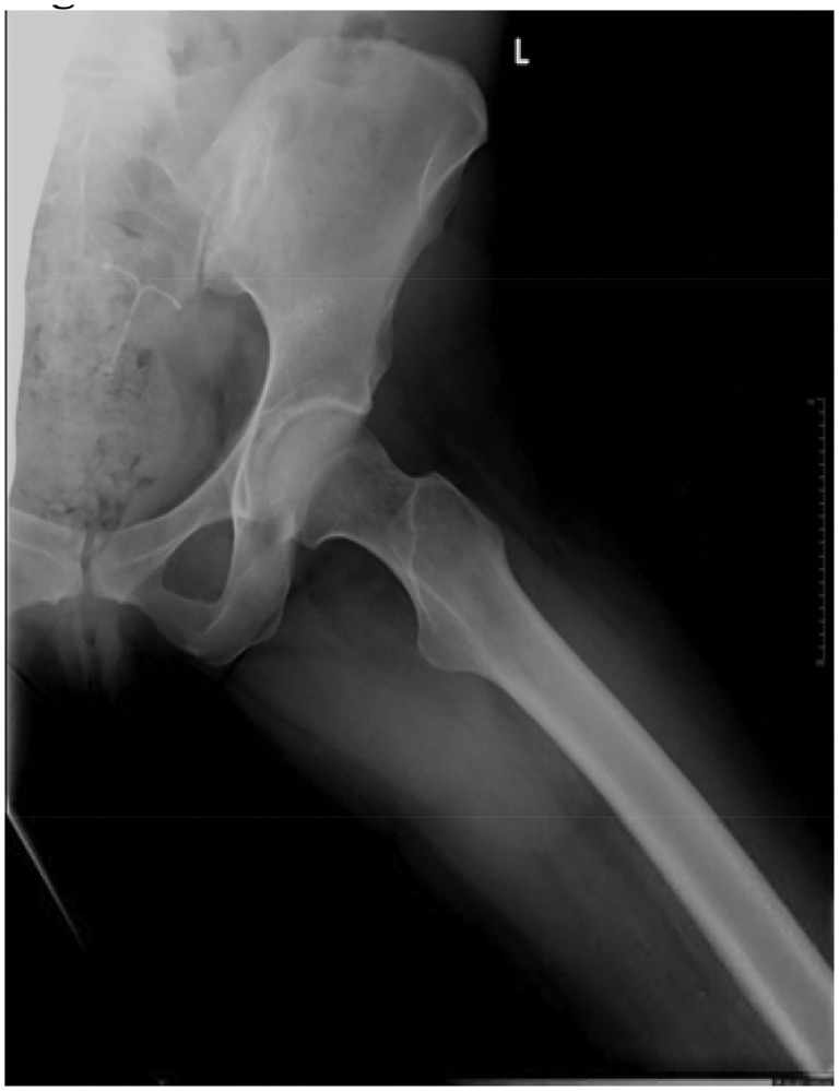
This report presents a case of a 37-year-old female with a history of hip pain. Magnetic resonance arthrography revealed loose bodies within the joint and synovial hypertrophy indicative of synovial chondromatosis (SC). Hip arthroscopy revealed free chondral bodies and focal villonodular synovial proliferation. The focal synovial proliferation was excised, a total synovectomy performed, and all cartilaginous free bodies removed. A post-operative histological examination of the removed nodular mass and synovium yielded evidence of both SC and pigmented villonodular synovitis (PVNS). A 1-year post-operative clinical examination showed marked clinical improvement and no signs of recurrence on MR images. Despite the clinical similarities, PVNS and SC are two distinct conditions that, to our knowledge, have never been reported as simultaneously occurring in a hip joint. The simultaneous presence of both pathologies may suggest a common origin of synovial metaplasia.
Figures


References
-
- Adelani MA, Wupperman RM, Holt GE.. Benign synovial disorders. J Am Acad Orthop Surg 2008; 16: 268–75. - PubMed
-
- Yoon PW, Yoo JJ, Koo KH. et al. Joint space widening in synovial chondromatosis of the hip. J Bone Joint Surg Am 2011; 93: 303–10. - PubMed
-
- Startzman A, Collins D, Carreira D.. A systematic literature review of synovial chondromatosis and pigmented villonodular synovitis of the hip. Phys Sportsmed 2016; 44: 425–31. - PubMed
Publication types
LinkOut - more resources
Full Text Sources
