. 2019:1899:211-227.
doi: 10.1007/978-1-4939-8938-6_15.

Clinical and Functional Evaluation of Ocular Inflammatory Disease Using the Model of Experimental Autoimmune Uveitis

Affiliations

Clinical and Functional Evaluation of Ocular Inflammatory Disease Using the Model of Experimental Autoimmune Uveitis

Jun Chen et al. Methods Mol Biol. 2019.

Abstract

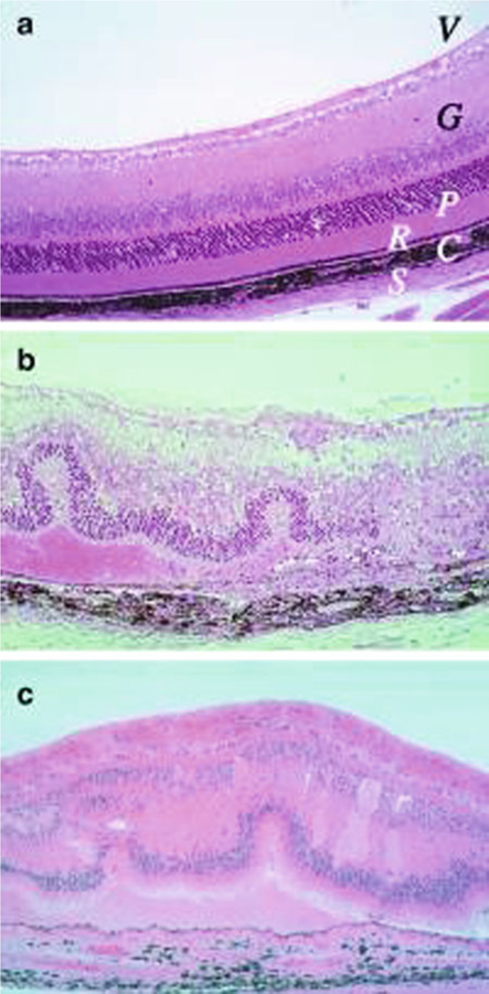
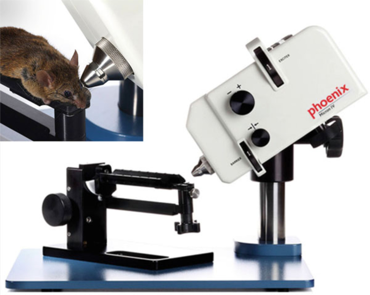
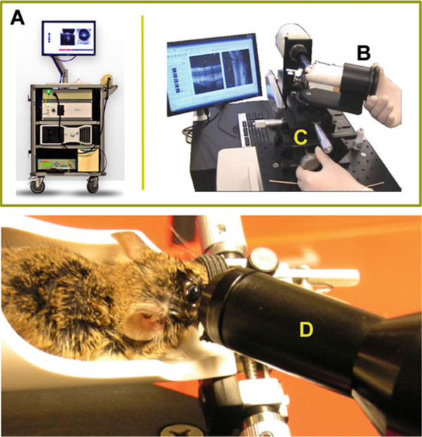
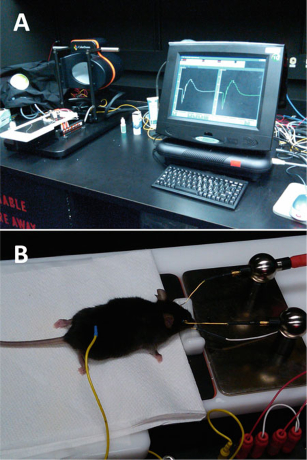
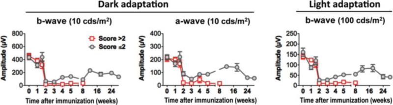
Non-infections uveitis in humans is an autoimmune disease of the retina and uvea that can be blinding if untreated. Its laboratory equivalent is experimental autoimmune uveitis (EAU) induced in susceptible rodents by immunization with retinal antigens and described elsewhere in this series (Agarwal et al., Methods Mol Biol, 900:443-469, 2012). Evaluation and quantitation of the disease is usually performed by fundus examination and/or histopathology, which provide limited information on structural and no information on functional changes as disease progresses. Here, we describe methods for systematic evaluation of disease using noninvasive clinical assessments by fundus examination and photography, optical coherence tomography, and functional evaluation by electroretinography, which are then compared to histopathology. Using these methodologies, we demonstrate that clinical variants of disease can be accurately evaluated both clinically and functionally, facilitating longitudinal follow-up and providing information that cannot be obtained by fundoscopy and histology alone. These methodologies can be useful to obtain additional information and to evaluate effects of therapeutic modalities under investigation.

Keywords: Autoimmunity; EAU; Electroretinography; Fundoscopy; Histology; IRBP; Mouse; Optical coherence tomography; S-Ag; T cells; Tolerance; Uveitis.


Figures

Fig. 1
Fig. 1
Histopathology of mouse EAU compared with human uveitis. Eyes were collected from B10.RIII mice before (a) and 21 days after uveitogenic immunization with IRBP (b). Note disorganized retinal architecture and damage to ganglion and photoreceptor cell layers, retinal folds, subretinal hemorrhage, vasculitis, focal damage to the retinal pigment epithelium, and choroiditis. Uveitis in the patient with ocular sarcoidosis (c). Note gross similarity in pathological picture between b and c (Photographs provided by Dr. Chi-Chao Chan, Laboratory of Immunology, National Eye Institute) (Reprinted from ref. 9)
Fig. 2
Fig. 2
Phoenix Micron II small animal retinal imaging system (Phoenix Research Laboratories, Inc). The apparatus is comprised of a base system that incorporates a host computer as well as a Phoenix StreamPix 5-Single camera and rodent imaging holder. Photographs are reproduced from the website of www.kellogg.umich.edu with permission
Fig. 3
Fig. 3
Bioptigen Spectral Domain Ophthalmic Imaging System (Bioptigen, Inc., Durham, NC). The apparatus consists of a base system (A), an animal imaging mount (B), and rodent alignment stage (C), which houses a SD-OCT probe (D). The base system incorporates a host computer, 840 nm OCT engine with reference arm attachment, and the probe. The SD-OCT scanner is encased in the animal imaging mount, which allows forward and backward adjustment of the probe. The InVivoVue Clinic software enables the creation, display, loading, and saving of OCT image files. The rodent alignment system contains an X-(micrometer), Y- (scissor jack), and Z-translators along with stereotactic rotational cassette (for holding the mouse) within a bushing and platform base. The entire device is attached to a slit-lamp base. Photographs are reproduced from Bioptigen, Inc., with permission
Fig. 4
Fig. 4
Espion E2 ERG recording system (Diagnosys LLC). The device is composed of a base system that incorporates a touchscreen PC configuration as well as an ERG dome and mouse Table (A). Mouse is placed on an animal table and connected to electrodes for ERG recording (B)
Fig. 5
Fig. 5
Fundus images of EAU in B10.RIII mice. Eyes were photographed with a Micron II fundus imaging system during the acute phase of disease (day 13–21), showing a range of disease severity scores that parallel the histology scores in Fig. 7
Fig. 6
Fig. 6
Comparison of the clinical course and disease severity in monophasic vs. chronic EAU by fundus examination and OCT imaging. EAU was induced in B10.RIII mice by active immunization with IRBP161–180 in adjuvant. Left panel, disease scores of EAU at different stages of disease were evaluated using an adapted fundus microscope. Right panel, B-scan OCT of the retina was evaluated at the indicated time points using a Bioptigen SD-OCT imaging system. Retinal thickness was measured and averaged from OCT images of the retina (adapted from ref. 27) (See Note 7)
Fig. 7
Fig. 7
Histopathology of EAU in the B10.RIII mouse. Eyes were collected from B10.RIII mice 21 days after uveitogenic immunization with IRBP, representing a range of disease scores (Figure is reprinted from ref. 26)
Fig. 8
Fig. 8
Comparison of fundus photography, OCT, and histology for the evaluation of the acute/monophasic and the chronic forms of EAU. Retinal lesions were visualized using Micron-II fundus imaging and Bioptigen SD-OCT imaging systems, and followed by histological examination of (a) monophasic, and (b) chronic forms of EAU. Note all images are of the same eye. Note engorged blood vessels and peri-vascular exudates (green arrow) in ganglion cell layer (GCL) and inner plexiform layer (IPL), vitreal and subretinal hemorrhages (red arrow, dark area) visible in all retinal layers and corresponding to the same lesions in the fundus image and in OCT B-scan cross-sections and choroidal inflammation (yellow arrow) in retinal pigment epithelium (RPE) and choroid (CH) (Figure is modified from refs. 14, 27) (See Notes 1–7)
Fig. 9
Fig. 9
Kinetics of dark- and light-adapted ERG responses in mouse model of EAU. Mice were monitored and followed up at the indicative time points by ERG. Amplitude of dark- and light-adapted ERGs was recorded and analyzed (adapted from ref. 27) (See Note 7)

References

    1. Streilein JW (2003) Ocular immune privilege: the eye takes a dim but practical view of immunity and inflammation. J Leukoc Biol 74:179–185 - PubMed
    1. Caspi RR, Roberge FG, Nussenblatt RB (1987) Organ-resident, nonlymphoid cells suppress proliferation of autoimmune T-helper lymphocytes. Science 237:1029–1032 - PubMed
    1. Stein-Streilein J (2008) Immune regulation and the eye. Trends Immunol 29:548–554 - PubMed
    1. Taylor AW (2007) Ocular immunosuppressive microenvironment. Chem Immunol Allergy 92:71–85 - PubMed
    1. Zhou R, Horai R, Silver PB, Mattapallil MJ, Zarate-Blades CR, Chong WP, Chen J, Rigden RC, Villasmil R, Caspi RR (2012) The living eye “disarms” uncommitted autoreactive T cells by converting them to Foxp3(+) regulatory cells following local antigen recognition. J Immunol 188:1742–1750 - PMC - PubMed