. 2019 Mar 15;30(6):766-777.
doi: 10.1091/mbc.E18-07-0462. Epub 2019 Jan 16.

GRASP depletion-mediated Golgi destruction decreases cell adhesion and migration via the reduction of α5β1 integrin

Affiliations

GRASP depletion-mediated Golgi destruction decreases cell adhesion and migration via the reduction of α5β1 integrin

Erpan Ahat et al. Mol Biol Cell. .

Abstract

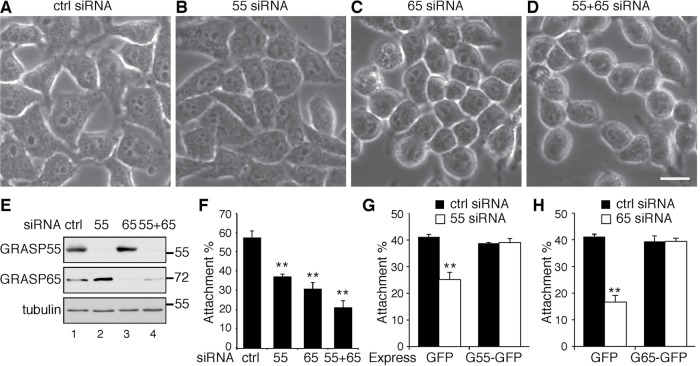
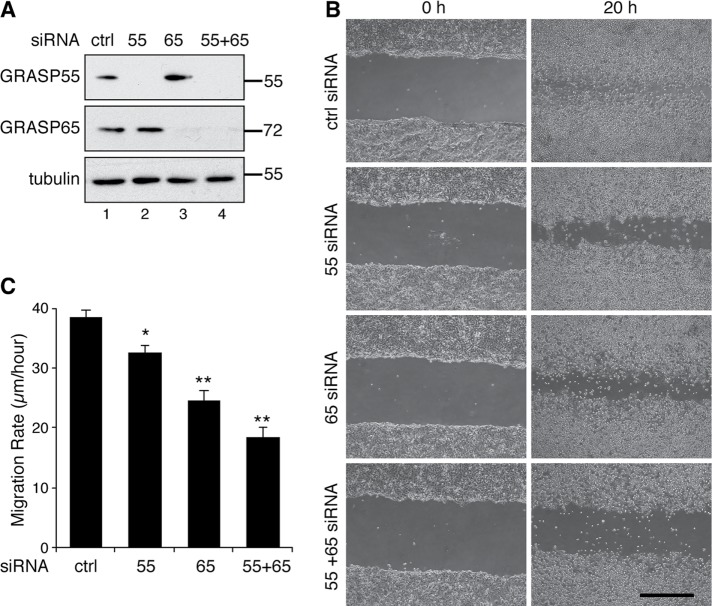
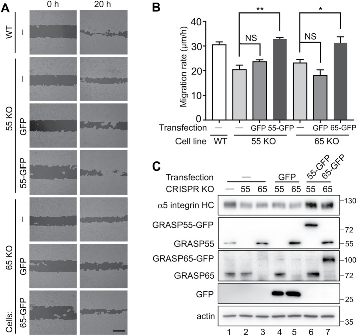
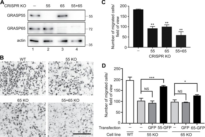
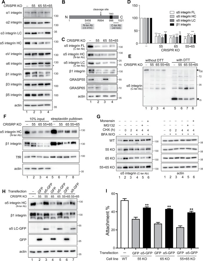
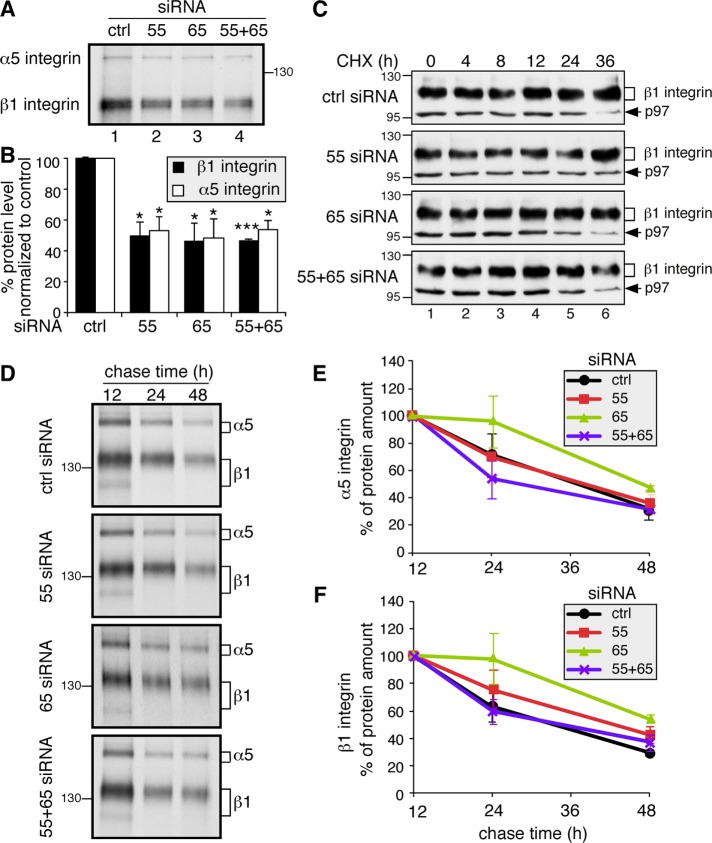
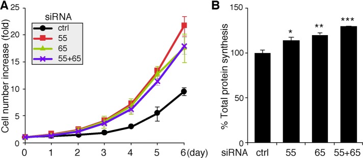
The Golgi apparatus is a membrane-bound organelle that serves as the center for trafficking and processing of proteins and lipids. To perform these functions, the Golgi forms a multilayer stacked structure held by GRASP55 and GRASP65 trans-oligomers and perhaps their binding partners. Depletion of GRASP proteins disrupts Golgi stack formation and impairs critical functions of the Golgi, such as accurate protein glycosylation and sorting. However, how Golgi destruction affects other cellular activities is so far unknown. Here, we report that depletion of GRASP proteins reduces cell attachment and migration. Interestingly, GRASP depletion reduces the protein level of α5β1 integrin, the major cell adhesion molecule at the surface of HeLa and MDA-MB-231 cells, due to decreased integrin protein synthesis. GRASP depletion also increases cell growth and total protein synthesis. These new findings enrich our understanding on the role of the Golgi in cell physiology and provide a potential target for treating protein-trafficking disorders.


Figures

FIGURE 1:
FIGURE 1:
GRASP depletion reduces cell attachment. (A–D) Phase-contrast images of HeLa cells transfected with indicated siRNAs. Scale bar: 20 µm. Note that GRASP-depleted cells are generally rounder than control siRNA-treated cells. (E) Western blots of HeLa cells transfected with indicated siRNAs. GRASP55 and GRASP65 were effectively depleted. (F) Cell attachment was reduced by GRASP depletion. A total of 3 × 10⁵ control (ctrl) or GRASP siRNA-treated cells were seeded on fibronectin-coated plates and incubated in serum-free medium for 30 min. After the removal of unbound cells, the number of attached cells was counted. Results are presented as mean ± SEM; statistical analysis was performed by comparison with control siRNA-treated cells using Student’s t test. **, p < 0.01. (G) The reduced attachment of GRASP55-depleted cells was rescued by the expression of GRASP55-GFP but not GFP. (H) The reduced attachment of GRASP65-depleted cells was rescued by expressing GRASP65-GFP.
FIGURE 2:
FIGURE 2:
GRASP depletion reduces cell migration. (A) Western blots of MDA-MB-231 cells transfected with the indicated siRNAs. GRASP55 and GRASP65 were effectively depleted. (B) GRASP depletion reduces MDA-MB-231 cell migration. Cells transfected with indicated siRNAs were analyzed in a wound-healing assay. Images were taken at 0 h and 20 h after scratching. Scale bar: 500 µm. (C) Quantitation of the migration rate in B. Statistical analysis was performed by comparison with control siRNA-treated cells using Student’s t test. *, p < 0.05; **, p < 0.01.
FIGURE 3:
FIGURE 3:
GRASP expression rescues the cell migration defects in GRASP-KO cells. (A) GRASP expression rescues the decreased cell migration in GRASP-KO HeLa cells. Cells transfected with indicated constructs were tested in a wound-healing assay, and images were processed by WimScratch: Wound Healing Assay Image Analysis Solution (Release 4.0). Images were taken at 0 and 20 h after scratching. Scale bar: 500 µm. (B) Quantitation of A. The reduced migration of GRASP55-KO and GRASP65-KO cells was rescued by expressing GRASP55-GFP and GRASP65-GFP, respectively, but not by GFP alone. Results are presented as mean ± SEM; statistical analysis was performed by comparison with WT control (ctrl) using Student’s t test. *, p < 0.05; **, p < 0.01. (C) Western blot of HeLa cells transfected with indicated constructs. α5-Integrin heavy chain (α5 integrin HC), GRASP55, GRASP65, GFP, and actin were blotted. The reduced protein levels of α5 integrin in GRASP55-KO and GRASP65-KO cells were rescued by expressing GRASP55-GFP or GRASP65-GFP, respectively, but not by GFP alone.
FIGURE 4:
FIGURE 4:
GRASP depletion reduces cell migration and invasion. (A) Western blots of WT and GRASP-KO HeLa cells for GRASP55, GRASP65, and actin. (B) GRASP depletion reduces cell migration and invasion. Indicated WT and GRASP-KO HeLa cells were analyzed in a Transwell assay. Images were taken after a 20-h migration. Scale bar: 500 µm. (C) Quantitation of the migration rate in B. Results are presented as mean ± SEM; statistical analysis was performed by comparison with WT cells using Student’s t test. (D) GRASP expression rescues the decreased cell migration in GRASP-KO cells. GRASP55-KO or GRASP65-KO cells transfected with indicated constructs were analyzed by a Transwell assay. Example images are shown in Supplemental Figure 3. Note that defects in cell invasion in GRASP55-KO and GRASP65-KO cells were rescued by the expression of GRASP55-GFP and GRASP65-GFP, respectively, but not by GFP alone. Results are presented as mean ± SEM; statistical analysis was performed by comparison with WT cells (ctrl) using Student’s t test. *, p < 0.05; **, p < 0.01; ***, p < 0.001.
FIGURE 5:
FIGURE 5:
GRASP knockout reduces α5β1-integrin protein level. (A) GRASP knockout reduces α5β1 level but has only modest effects on other integrins. Western blots of WT and GRASP-KO HeLa cells for indicated integrins and actin. Note that the protein levels of α1, αV, and α6 did not change after GRASP depletion in contrast to the robust reduction of α5 and β1 integrins. (B) Schematic α5-integrin domain structure and epitopes of indicated antibodies. (C) GRASP knockout reduces the α5β1-integrin level. The full-length (FL), heavy-chain (HC), and light-chain (LC) forms of α5 integrin, β1 integrin, GRASP55, GRASP65, and actin were blotted. Note the reduced protein level of α5 integrin on all three blots as well as β1 integrin compared with that in WT cells. (D) Quantitation of C normalized with actin. Statistical analysis was performed by comparison with WT cells. (E) Western blot of α5 integrin on nonreducing (without DTT) and reducing gels (with DTT). Arrow and arrowhead indicate full length and light chain, respectively. (F) GRASP knockout reduces α5β1-integrin level at the cell surface. GRASP-KO cells were surface biotinylated, this was followed by streptavidin pull-down and Western blotting of α5 integrin, β1 integrin, TfR, and actin. Note that only the highly glycosylated forms of integrins were pulled down; this was clearer with β1 integrin (top band). (G) GRASP knockout accelerates α5-integrin maturation. WT and GRASP-KO HeLa cells were pretreated with BFA for 2 h and washed out and then released into CHX for 0, 2, or 4 h or CHX for 4 h with MG132 or monensin. As GRASP-KO cells have a lower level of integrin level, we exposed those gels longer, so all cell lines had a similar signal at the 0 time point to start with, and the reduction of the protein was assessed over time. (H) Western blot of indicated HeLa cells transfected with α5 integrin-GFP or GFP. α5-Integrin heavy chain (α5-integrin HC), β1 integrin, GFP, and actin were blotted. The reduced protein levels of α5 and β1 integrins in GRASP-KO cells were rescued by expressing α5 integrin-GFP, but not by GFP alone. (I) α5-Integrin expression rescues the decreased cell attachment of GRASP-KO cells. Cells transfected with indicated constructs were analyzed in an attachment assay, as described in Figure 1. The quantitation results are shown. **, p < 0.01; ***, p < 0.001.
FIGURE 6:
FIGURE 6:
GRASP depletion reduces α5- and β1-integrin synthesis but has no effect on their turnover. (A) GRASP depletion reduces protein synthesis of α5 and β1 integrin. An equal number of HeLa cells transfected with indicated siRNAs were labeled with Trans 35S-Label [35S] for 1 h. Immunoprecipitated α5β1 integrin was analyzed by gel electrophoresis and autoradiography. The protein synthesis rates of both α5 and β1 integrin decreased in GRASP-depleted cells. (B) Quantitation of A. Statistical analysis was performed by comparison with control (ctrl) siRNA-treated cells. *, p < 0.05; ***, p < 0.001. (C) Western blot of GRASP-depleted HeLa cells treated with CHX for the indicated times. At 72 h posttransfection with the indicated siRNA, cells were treated with 100 µM CHX for 0, 4, 8, 12, 24, and 36 h; lysed; and analyzed by Western blot for β1 integrin and p97 on the same gel. As GRASP knockdown cells have a lower level of integrins, we exposed those gels longer, so all cell lines had a similar signal at the 0 time point to start with, and the reduction of the protein was assessed over time. (D) GRASP depletion does not increase α5- and β1-integrin degradation. HeLa cells transfected with indicated siRNAs were labeled with Trans 35S-Label [35S] for 1 h and chased for 12, 24, and 48 h. Immunoprecipitated α5β1 integrins were analyzed by SDS–PAGE and autoradiography. (E) Quantification of α5 integrin in D. Note that there is no significant difference in the degradation rate of α5 integrin between control siRNA-treated and GRASP-depleted cells. (F) Quantification of β1 integrin in D. There is no significant difference in the degradation rate of β1 integrin between control siRNA-treated and GRASP-depleted cells.
FIGURE 7:
FIGURE 7:
Depletion of GRASP55 and/or GRASP65 enhances cell growth and total protein synthesis. (A) Growth rate of HeLa cells transfected with the indicated siRNAs, as measured using crystal violet staining. The measurement began at 48 h after transfection. (B) Total protein synthesis is enhanced in GRASP-depleted cells. HeLa cells transfected with indicated siRNAs were labeled with Trans 35S-Label [35S] for 1 h. Equal amounts of total proteins were precipitated with TCA, and [35S]methionine/cysteine incorporation was assessed by scintillation counting. Statistical analysis was performed by normalization and comparison with control (ctrl) siRNA-treated cells using Student’s t test. *, p < 0.05; **, p < 0.01; ***, p < 0.001.

References

    1. Aridor M, Balch WE. (1999). Integration of endoplasmic reticulum signaling in health and disease. Nat Med , 745–751. - PubMed
    1. Barr FA, Puype M, Vandekerckhove J, Warren G. (1997). GRASP65, a protein involved in the stacking of Golgi cisternae. Cell , 253–262. - PubMed
    1. Bekier ME, 2nd, Wang L, Li J, Huang H, Tang D, Zhang X, Wang Y. (2017). Knockout of the Golgi stacking proteins GRASP55 and GRASP65 impairs Golgi structure and function. Mol Biol Cell , 2833–2842. - PMC - PubMed
    1. Bergman M, Joukov V, Virtanen I, Alitalo K. (1995). Overexpressed Csk tyrosine kinase is localized in focal adhesions, causes reorganization of alpha v beta 5 integrin, and interferes with HeLa cell spreading. Mol Cell Biol , 711–722. - PMC - PubMed
    1. Bizzaro N, Pasini P, Ghirardello A, Finco B. (1999). High anti-Golgi autoantibody levels: an early sign of autoimmune disease? Clin Rheumatol , 346–348. - PubMed

Publication types

LinkOut - more resources