. 2019 Jan 16;14(1):e0209414.
doi: 10.1371/journal.pone.0209414. eCollection 2019.

Characterisation of ovine lymphatic vessels in fresh specimens

Affiliations

Characterisation of ovine lymphatic vessels in fresh specimens

Hung-Hsun Yen et al. PLoS One. .

Abstract

Background and aim: The development and use of experimental models using lymphatic cannulation techniques have been hampered by the lack of high-quality colour imaging of lymphatic vessels in situ. Most descriptions of lymphatic anatomy in sheep have historically depended on schematic diagrams due to limitations in the ability to publish colour images of the lymphatic vessels with decent resolution. The aim of this work was to encourage more widespread use of the ovine cannulation model by providing clear photographic images identifying the location and anatomical layout of some major lymphatic ducts and their in situ relationship to surrounding tissues.

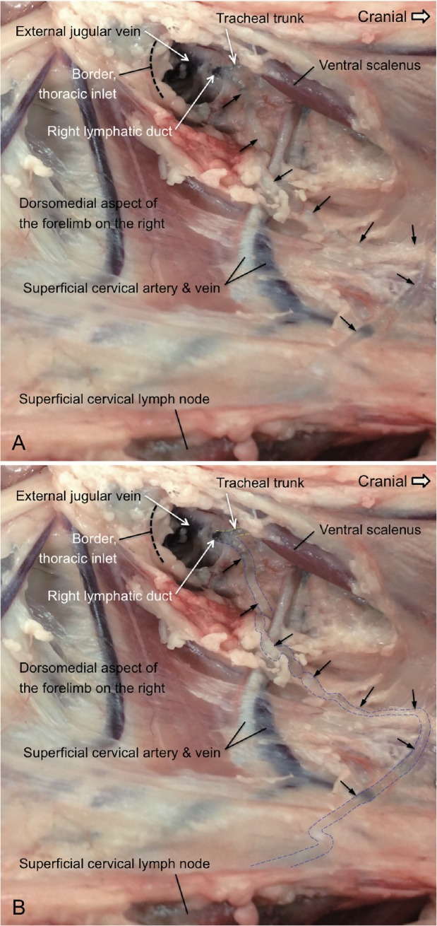
Methods: The cadavers of the sheep were collected after they had been euthanized at the end of animal trials not associated with this study. The lymphatics were dissected and exposed to show their appearance in the surrounding tissues and their relationship to other organs. Patent Blue was used to locate lymphatic vessels in exploratory preparations. However, in order to present the natural appearance of the vessels, we used minimal dissection and dye was not used for the photographed examples. Instead, we have indicated the course of the vessels with lines where their position is less clear.

Results and conclusion: In this paper, we have used sheep specimens as examples to show characteristic images of lymphatic vessels. The images of in situ lymphatics and lymph nodes combined with schematic summaries provide a concise illustration of the lymphatic drainage scheme in sheep.


Conflict of interest statement

The authors have declared that no competing interests exist.

Figures

Fig 1
Fig 1. Survey diagram of the lymph nodes of sheep.
Two schematic images illustrate the principal lymphatic vessels and the lymph nodes of sheep. I. Images showing the tracheal trunks are available in the previous report by Yen at al (Yen et al., 2006) [7]. II. The image showing the junction of the thoracic duct and the efferent lymphatic of the superficial cervical lymph node(s) on the left can be found in the previous report by Yen at al (Yen et al., 2009) [8]. III. A segment of the thoracic duct dorsal to the aorta and the caudal mediastinal lymph node in the caudal thoracic cavity on the right are illustrated in Fig 2. IV. Images showing the efferent lymphatic vessels of the mammary glands are available in the previous reference by Yen et al (Yen et al., 2016) [9]. V. The right lymphatic duct, the tracheal trunk and the efferent lymphatic of the superficial cervical lymph node(s) on the right are depicted in Fig 3. VI. The visceral, hepatic and intestinal trunks are depicted in Fig 4. VII. The afferent and efferent lymphatic vessels of a hepatic lymph node are depicted in Fig 5. VIII. The efferent lymphatic of the ileocolic lymph nodes and the jejunal trunk are shown in Fig 6. 1&1’: left and right parotid lymph nodes; 2&2’: left and right mandibular lymph nodes; 3&3’: left and right medial retropharyngeal lymph nodes; 4&4’: left and right lateral retropharyngeal lymph nodes; 5: cranial deep cervical lymph nodes; 6: middle deep cervical lymph nodes; 7: caudal deep cervical lymph nodes; 8&8’: left and right accessary superficial cervical lymph nodes; 9&9’: left and right superficial cervical lymph nodes; 10: costocervical lymph nodes; 11: cranial mediastinal lymph node(s); 12: middle mediastinal lymph node(s); 13: intercostal lymph nodes; 14: thoracic aortic lymph nodes; 15: caudal mediastinal lymph nodes; 16: tracheobronchial lymph center; 17: pulmonary lymph nodes (left and right); 18: cranial and caudal sternal lymph nodes; 19: costoaxillary lymph nodes; 20&20’: left and right axillary lymph nodes; 21&21’: left and right accessary axillary lymph nodes (inconstant); 22: lumbar aortic lymph nodes; 23: medial iliac lymph node(s); 24: sacral lymph nodes; 25&25’: left and right iliofemoral lymph nodes (inconstant); 26&26’: left and right subiliac (or prefemoral) lymph nodes; 27&27’: left and right mammary (or superficial inguinal) lymph nodes; 28&28’: left and right sciatic (or ischiadic) lymph nodes; 29&29’: left and right anorectal lymph nodes; 30: left and right popliteal lymph nodes; 31: atrial lymph nodes; 32: reticular lymph nodes; 33: omasal lymph nodes; 34: dorsal abomasal lymph nodes; 35: right ruminal lymph nodes; 36: hepatic lymph nodes; 37: renal lymph nodes; 38: celiac and cranial mesenteric lymph nodes (inconstant); 39: pancreaticoduodenal lymph nodes; 40: jejunal lymph nodes; 41: ileocolic lymph nodes; 42: colic nodes; 43: caudal mesenteric lymph nodes (inconstant); a: trachea; b&b’: left and right tracheal trunks; c: thoracic duct; d&d’: left and right external jugular veins; e: left subclavian vein; f: cranial vena cava; g: cisterna chyli; h: lumbar trunk; i: aorta; j: esophagus; k: diaphragm; l: right lymphatic duct; m: visceral trunk; n: gastric trunk; o: hepatic trunk; p: intestinal trunk; q: jejunal trunk.
Fig 2
Fig 2. The dissection of an ovine thoracic duct in the caudal thorax on the right.
A. The locations of the thoracic duct, the caudal mediastinal lymph node and a short segment of its efferent lymphatic on the right side of the thorax are depicted in this image. The thoracic duct was located dorsal to the aorta right after it passed through the diaphragm and entered the thoracic cavity. In most animals, the thoracic duct is to some extent shifted to the right in the caudal thorax. An efferent lymphatic vessel of the caudal mediastinal lymph node embedded in fat, departed at the cranial end of the lymph node and led dorsally to the thoracic duct. The setting for the contrast and brightness of this image is for the presentation of the lymphatics rather than the liver and the diaphragm. The depth differences between the thoracic duct and the livers in this image are huge; therefore the liver and the diaphragm look dark. This figure is positioned with cranial to the right. B. The photograph is the same as Fig 2A. In this figure, the yellow dotted lines indicate the location of three segments of the thoracic duct. These segments are not embedded in the adipose tissues. The blue dotted lines mark the visible part of the efferent lymphatic vessel of the caudal mediastinal lymph node.
Fig 3
Fig 3. The dissection of an ovine right lymphatic duct.
A. The locations of the right lymphatic duct at its point of entry into the external jugular vein on the right are depicted in this image. The efferent lymphatic vessel of the superficial cervical lymph node(s) on the right coursed medial to the superficial cervical artery and vein toward the thoracic inlet. This lymphatic merged with the tracheal trunk at the location just cranial to the thoracic inlet to become the right lymphatic duct. The black arrows depict the efferent lymphatic vessel of the superficial cervical lymph node(s). This figure is positioned with cranial to the right and shows the dorsal aspect of the caudal cervical and axillary regions medial to the forelimb on the right. B: This photograph is the same as Fig 3A. In this image, the yellow dotted lines mark the location of the tracheal trunk and the blue dotted lines indicate the course of the efferent lymphatic of the superficial cervical lymph node. Ventral scalenus: the ventral scalenus muscle.
Fig 4
Fig 4. The dissection of an ovine visceral trunk, hepatic trunk and intestinal trunk.
The locations of the visceral trunk and the confluence of the hepatic and intestinal trunks are present in this image. Lymph in the intestinal trunk contains abundant lipids following normal feeding and is shown by the cloudy appearance. Lymph from the intestinal trunk drains into the visceral trunk and thus the visceral trunk also presents the “milky” color. As depicted in Fig 1B, the visceral trunk collects lymph from many major organs in the abdominal cavity. Lymph from the gastric trunk, hepatic trunk and intestinal trunk all streams into the visceral trunk. The visceral trunk enters the lumbar trunk next to its junction with the cisterna chyli. This figure is positioned with cranial to the right and shows the ventrolateral aspect of the region caudal to the liver on the right. G: gloves.
Fig 5
Fig 5. The dissection of one ovine hepatic lymph node and its afferent and efferent lymphatic vessels.
The location of a hepatic lymph node at the junction of the portal vein and pancreas is shown in this image. Multiple afferent lymphatic vessels coming from the liver alongside the portal vein are entering this lymph node. There was an efferent lymphatic, a branch of the hepatic trunk, leaving this lymph node. This image demonstrates the usual number of afferent and efferent lymphatic vessels of a lymph node. This figure is positioned with cranial to the left and caudal to the right and shows the ventrolateral aspect of the tissues between the pancreas and the liver on the right hand side. Portal vein: the wall of the portal vein is shown lying beneath the clear afferent lymphatics.
Fig 6
Fig 6. The dissection of an ovine jejunal lymphatic trunk and the efferent lymphatic of the ileocolic lymph node(s).
A. The lymph nodes and lymphatics exposed in the initial dissection of ovine mesentery. The ileocolic lymph node is shown at the end of the cecum (cecocolic junction). The large, discrete terminal jejunal lymph node indicated in the image has previously been referred to as the ileocecal lymph node and drains the terminal ileum [13]. B. The locations of the jejunal lymphatic trunk which is a branch of the intestinal trunk, and an efferent lymphatic of the ileocolic lymph node are depicted in this image. This efferent lymphatic of the ileocolic lymph node(s) is a segment of the lymphatic that is located proximal to its join to the intestinal trunk. The jejunal lymphatic shows the usual “milky” appearance due to the presence of chyle in its lumen. The jejunal trunk collects lymph draining the jejunal lymph nodes. The jejunal trunk is associated with the mesentery that lies on the colon. This figure is positioned with cranial to the right. C: This photograph is the same as Fig 6B. In this image, the yellow dotted line marks a segment of the efferent lymphatic of the ileocolic lymph nodes. The efferent lymphatic of the ileocolic lymph node and the jejunal lymphatic trunk are also presented in the video (S1 File) attached.

Similar articles

Cited by

References

    1. Washington EA, Barber SR, Murray CM, Davies HMS, Kimpton WG, Yen HH. Lymphatic cannulation models in sheep: Recent advances for immunological and biomedical research. J Immunol Methods. 2018;457:6–14. Epub 2018/04/07. 10.1016/j.jim.2018.03.011 . - DOI - PubMed
    1. Hein WR, Griebel PJ. A road less travelled: large animal models in immunological research. Nat Rev Immunol. 2003;3(1):79–84. 10.1038/nri977 . - DOI - PubMed
    1. Pearson LD, Simpson-Morgan MW, Morris B. Lymphopoiesis and lymphocyte recirculation in the sheep fetus. J Exp Med. 1976;143(1):167–86. - PMC - PubMed
    1. Scheerlinck JP, Gekas S, Yen HH, Edwards S, Pearse M, Coulter A, et al. Local immune responses following nasal delivery of an adjuvanted influenza vaccine. Vaccine. 2006;24(18):3929–36. 10.1016/j.vaccine.2006.02.032 . - DOI - PubMed
    1. Burke ML, de Veer M, Pleasance J, Neeland M, Elhay M, Harrison P, et al. Innate immune pathways in afferent lymph following vaccination with poly(I:C)-containing liposomes. Innate Immun. 2014;20(5):501–10. 10.1177/1753425913501213 . - DOI - PubMed