. 2019 Jan 15;20(2):326.
doi: 10.3390/ijms20020326.

Osteochondral Tissue Regeneration Using a Tyramine-Modified Bilayered PLGA Scaffold Combined with Articular Chondrocytes in a Porcine Model

Affiliations

Osteochondral Tissue Regeneration Using a Tyramine-Modified Bilayered PLGA Scaffold Combined with Articular Chondrocytes in a Porcine Model

Tzu-Hsiang Lin et al. Int J Mol Sci. .

Abstract

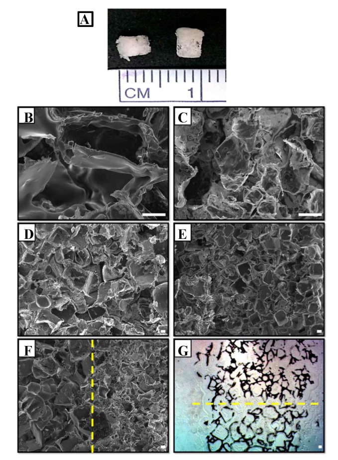
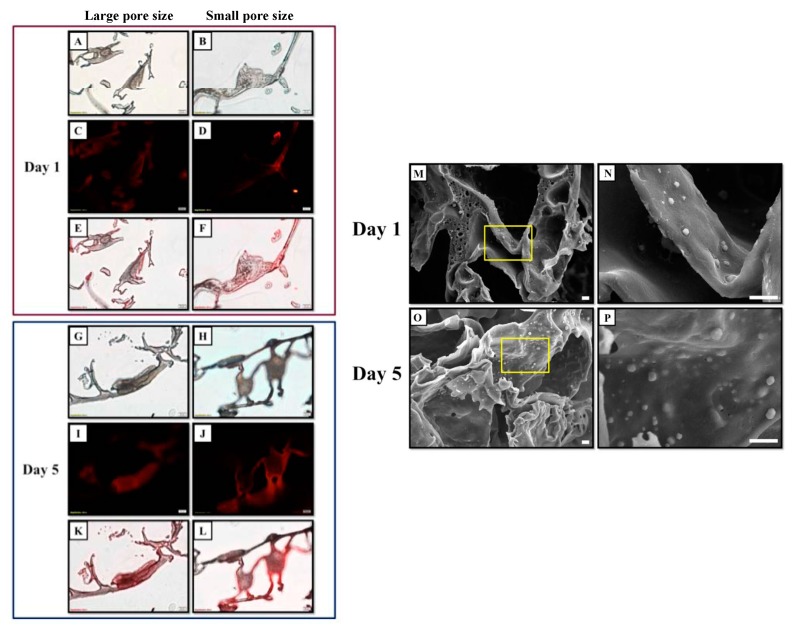
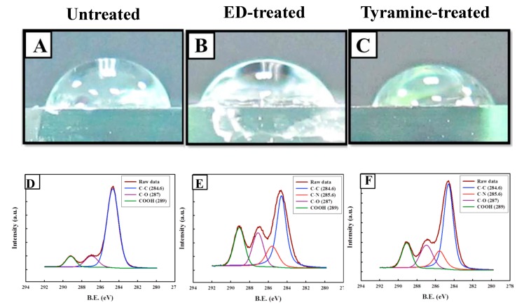
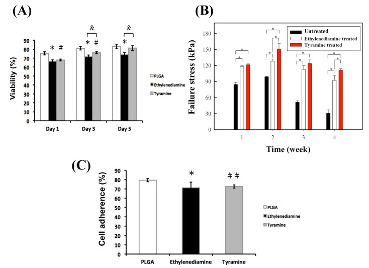
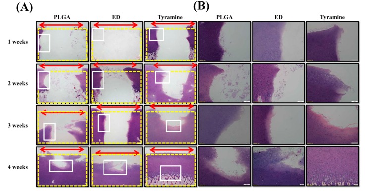
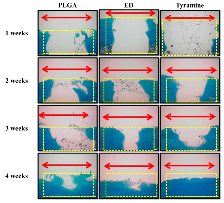
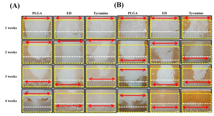
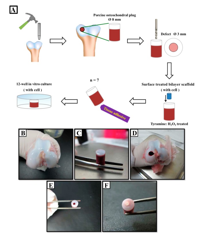
Repairing damaged articular cartilage is challenging due to the limited regenerative capacity of hyaline cartilage. In this study, we fabricated a bilayered poly (lactic-co-glycolic acid) (PLGA) scaffold with small (200⁻300 μm) and large (200⁻500 μm) pores by salt leaching to stimulate chondrocyte differentiation, cartilage formation, and endochondral ossification. The scaffold surface was treated with tyramine to promote scaffold integration into native tissue. Porcine chondrocytes retained a round shape during differentiation when grown on the small pore size scaffold, and had a fibroblast-like morphology during transdifferentiation in the large pore size scaffold after five days of culture. Tyramine-treated scaffolds with mixed pore sizes seeded with chondrocytes were pressed into three-mm porcine osteochondral defects; tyramine treatment enhanced the adhesion of the small pore size scaffold to osteochondral tissue and increased glycosaminoglycan and collagen type II (Col II) contents, while reducing collagen type X (Col X) production in the cartilage layer. Col X content was higher for scaffolds with a large pore size, which was accompanied by the enhanced generation of subchondral bone. Thus, chondrocytes seeded in tyramine-treated bilayered scaffolds with small and large pores in the upper and lower parts, respectively, can promote osteochondral regeneration and integration for articular cartilage repair.

Keywords: PLGA; bilayer; chondrocyte; osteochondral regeneration; tyramine.


Conflict of interest statement

The authors declare no conflict of interest.

Figures

Figure 1
Figure 1
SEM images of bilayered porous poly (lactic-co-glycolic acid) (PLGA) scaffolds. (A) Macroscopic view of the scaffold. (BG) Microscopic views of the scaffold: high magnification image of large pores (200×) (B); high magnification image of small pores (200×) (C); low magnification image of large pores (50×) (D); low magnification image of small pores (50×) (E); bilayered PLGA scaffold (50×) (F); and light micrograph of a frozen section of a bilayered PLGA scaffold (G). The yellow dotted line in (F) and (G) represents the boundary between the lower (large pore) and upper (small pore) layers. Scale bar: 100 μm.
Figure 2
Figure 2
Images of chondrocytes on PLGA scaffolds with different pores sizes after one and five days of culture. The cell membrane was labeled with red fluorescent dye. (A,B,G,H) Light micrographs. (C,D,I,J) Fluorescence micrographs. (E,F,K,L) Merged light and fluorescence micrographs. The red and blue boxes indicate the images of cells on PLGA scaffolds at day one and five, respectively. Scale bar: 20 μm. (MP) SEM images of cell-seeded PLGA scaffold with a small pore size: images of cells cultured on PLGA scaffolds on day one (M,N) and day five (O,P). The area enclosed by the yellow box is shown enlarged to the right. Scale bar: 10 μm.
Figure 3
Figure 3
Water contact angle and results of surface analysis by X-ray photoelectron spectroscopy (XPS) of untreated and surface-treated PLGA scaffolds. (A,D) Untreated scaffolds. (B,E) Ethylenediamine (ED)-treated scaffolds. (C,F) Tyramine-treated scaffolds.
Figure 4
Figure 4
(A) Cell viability. Values represent mean ± SD (n = 6). * p < 0.05 and # p < 0.05: ED-treated or tyramine-treated groups versus the untreated PLGA group, respectively. & p < 0.05: ED-treated group versus the tyramine-treated PLGA group. (B) Push-out test results for untreated and surface-treated PLGA scaffolds. Values represent mean ± SD (n = 4). * p < 0.05. (C) Cell adherence of chondrocytes cultured in untreated, ED- or tyramine-treated PLGA scaffolds at day one. Values represent mean ± SD (n = 4). * p < 0.05 and ## p < 0.01: ED- or tyramine-treated groups versus the untreated PLGA group, respectively.
Figure 5
Figure 5
Histological analysis of co-cultured bilayered PLGA scaffold and porcine osteochondral plug by hematoxylin and eosin staining. (A) Magnification: 40×; scale bar: 200 μm. (B) Magnification: 100×; scale bar: 100 μm. Red double-headed arrows (←→) indicate the defect area after tissue regeneration. Yellow dotted boxes indicate the defect area before tissue regeneration. White boxes indicate that the images of tissue regeneration area were amplified.
Figure 6
Figure 6
Histological analysis of co-cultured bilayered PLGA scaffold and porcine osteochondral plug by Alcian blue staining. Magnification: 40×; scale bar: 200 μm. Red double-headed arrows (←→) indicate the defect area after tissue regeneration. Yellow dotted boxes indicate the defect area before tissue regeneration.
Figure 7
Figure 7
(A,B) Immunohistochemical detection of collagen type X (Col X) (A) and collagen type II (Col II) (B) in a co-cultured bilayered PLGA scaffold and porcine osteochondral plug. Magnification: 40×; scale bar: 200 μm. Red double-headed arrows (←→) indicate the defect area after tissue regeneration. Yellow dotted boxes indicate the defect area before tissue regeneration. White dotted lines are used to distinguish the areas for cartilage and subchondral bone.
Figure 8
Figure 8
Fabrication of osteochondral host tissue plug and ex vivo co-culture of plug tissue and cell-seeded surface-treated scaffold. (A) Schematic illustration of porcine plug manufacture and co-culture of plug tissue and cell-seeded, surface-treated scaffold. Thick white arrows indicate the processes of osteochondral plug manufacture and the cell-seeded surface-treated scaffold implantation; thin black arrow indicates the implantation of cell-seeded surface-treated scaffold in porcine plug; the blue cylinder indicates the cell-seeded surface-treated scaffold. (B) Femoral condyles of porcine knee joints. (C) An osteochondral tissue plug was created using an eight-mm diameter hole driller. (D) Porcine femur after plug removal. (E) A three-mm defect was drilled into the osteochondral plug. (F) A three-mm cell-seeded bilayered PLGA scaffold was pressed into the defect.

Similar articles

Cited by

References

    1. Kocher M.S., Tucker R., Ganley T.J., Flynn J.M. Management of osteochondritis dissecans of the knee: Current concepts review. Am. J. Sports Med. 2006;34:1181–1191. doi: 10.1177/0363546506290127. - DOI - PubMed
    1. Shea K.G., Jacobs J.C., Jr., Carey J.L., Anderson A.F., Oxford J.T. Osteochondritis dissecans knee histology studies have variable findings and theories of etiology. Clin. Orthop. Relat. Res. 2013;471:1127–1136. doi: 10.1007/s11999-012-2619-6. - DOI - PMC - PubMed
    1. Versier G., Dubrana F., French Arthroscopy S. Treatment of knee cartilage defect in 2010. Orthop. Traumatol. Surg. Res. 2011;97:S140–S153. doi: 10.1016/j.otsr.2011.09.007. - DOI - PubMed
    1. Bobic V. Arthroscopic osteochondral autograft transplantation in anterior cruciate ligament reconstruction: A preliminary clinical study. Knee Surg. Sports Traumatol. Arthrosc. 1996;3:262–264. doi: 10.1007/BF01466630. - DOI - PubMed
    1. Hangody L., Kish G., Karpati Z., Udvarhelyi I., Szigeti I., Bely M. Mosaicplasty for the treatment of articular cartilage defects: Application in clinical practice. Orthopedics. 1998;21:751–756. doi: 10.3928/0147-7447-19980701-04. - DOI - PubMed

LinkOut - more resources