Review
. 2019 Jan 15;11(1):65.
doi: 10.3390/v11010065.

Neuroinflammation, Microglia, and Cell-Association during Prion Disease

Affiliations
Review

Neuroinflammation, Microglia, and Cell-Association during Prion Disease

James A Carroll et al. Viruses. .

Abstract

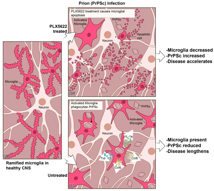
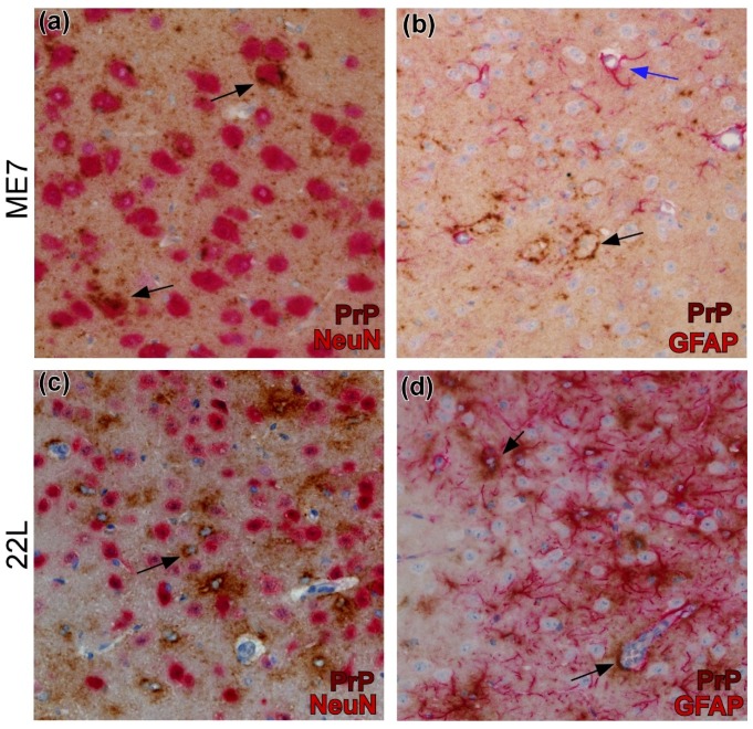
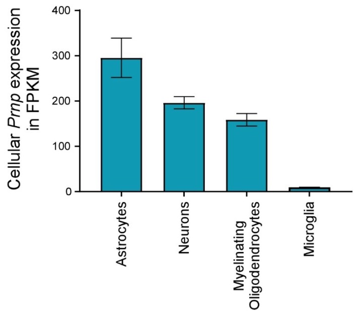
Prion disorders are transmissible diseases caused by a proteinaceous infectious agent that can infect the lymphatic and nervous systems. The clinical features of prion diseases can vary, but common hallmarks in the central nervous system (CNS) are deposition of abnormally folded protease-resistant prion protein (PrPres or PrPSc), astrogliosis, microgliosis, and neurodegeneration. Numerous proinflammatory effectors expressed by astrocytes and microglia are increased in the brain during prion infection, with many of them potentially damaging to neurons when chronically upregulated. Microglia are important first responders to foreign agents and damaged cells in the CNS, but these immune-like cells also serve many essential functions in the healthy CNS. Our current understanding is that microglia are beneficial during prion infection and critical to host defense against prion disease. Studies indicate that reduction of the microglial population accelerates disease and increases PrPSc burden in the CNS. Thus, microglia are unlikely to be a foci of prion propagation in the brain. In contrast, neurons and astrocytes are known to be involved in prion replication and spread. Moreover, certain astrocytes, such as A1 reactive astrocytes, have proven neurotoxic in other neurodegenerative diseases, and thus might also influence the progression of prion-associated neurodegeneration.

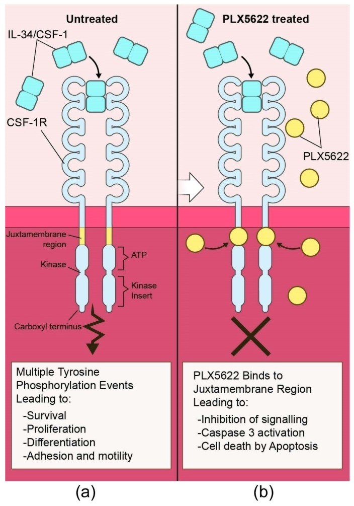
Keywords: PLX5622; astroglia; chemokine; cytokine; microglia; neuroinflammation; neuron; prion; scrapie.


Conflict of interest statement

The authors declare no conflict of interest.

Figures

Figure 1
Figure 1
Quantitative reverse transcription-polymerase chain reaction (qRT-PCR) analysis of expression of 84 proinflammatory genes in the brains of mice at 70 compared to 131 days after prion infection. Each black dot correlates to the average gene expression seen in a minimum of three mice. The orange box is the region containing proinflammatory genes that are statistically significant by t-test (p ≤ 0.05, the black dashed line) and greater than 2-fold increased (the blue dashed line) relative to mock infected control mice. The number and magnitude of the upregulated proinflammatory genes in the brain of prion infected mice intensifies as a function of time.
Figure 2
Figure 2
Comparison of the top 25 proinflammatory genes that are upregulated in the brain of clinical mice when infected with prion strains RML and 22L. The orange dashed line represents a one-to-one correlation in fold change of gene expression. Most of the changes are similar, thus close to the orange dotted line. There is a cluster of eight proinflammatory genes, blue circle, that are more highly altered with 22L infection, but the overall inflammation is comparable during the clinical phase of the disease regardless of prion strain.
Figure 3
Figure 3
Representation of the CSF-1 Receptor (CSF-1R) interaction with the tyrosine kinase inhibitor PLX5622. Under normal conditions (a) CSF-1R interacts with either CSF-1 or IL-34 to promote phosphorylation at a minimum of eight tyrosine residues within the cytoplasmic domains. These phosphorylation sites serve as docking points for many proteins and lead to downstream signaling events. Inhibition of CSF-1R with PLX5622 (b) causes a cessation of signaling through this receptor in microglia, which is critical for their survival and proliferation. Microglia are eliminated from the CNS by activation of Caspase 3, leading to death by apoptosis.
Figure 4
Figure 4
Illustration of prion disease with and without PLX5622 treatment to reduce microglia in the Central Nervous System (CNS). (a) Microglia in the healthy CNS typically have a ramified appearance as they surveil their environment. (b) When prion-infected mice are treated with PLX5622, microglia are reduced, the level of PrPSc increases, and the disease process is accelerated. (c) When prion-infected mice are untreated, more microglia are present to phagocytize PrPSc and to produce microglial specific proinflammatory effectors like TNF, CCL6, CCL8, and IL-1a. The presence of microglia reduces the PrPSc burden and lengthens the disease.
Figure 5
Figure 5
Use of dual-staining immunohistochemistry for detection of PrPSc from scrapie strains ME7 and 22L associated with neurons or astrocytes in brain. (a) PrPSc (brown) surrounds ME7-infected neurons (arrows) in amygdala that are detected by anti-NeuN (red). (b) In hypothalamus, ME7 PrPSc surrounds neuronal cell bodies (black arrow). Astrocytes detected by anti-Glial Fibrillary Acidic Protein (GFAP) (red) have no PrPSc and are not infected (blue arrow). (c) PrPSc (brown)-expressing 22L-infected cells (arrow) are distinct from NeuN-stained (red) neurons in thalamus. (d) PrPSc (brown) expressing 22L-infected cells (arrows) are associated with GFAP-stained (red) astrocytes in thalamus.
Figure 6
Figure 6
Cellular Prnp expression by various brain cells in healthy mice. RNA seq data of purified cells isolated from health mouse brain was acquired from the work of Zhang et al. and represented here in fragments per kilobase million (FPKM) for comparison [165]. The blue columns are the average of two replicates of pooled animals for each cell type and bars represent the standard deviation. Astrocytes express the most Prnp, while microglia express very little.

References

    1. Caughey B., Baron G.S., Chesebro B., Jeffrey M. Getting a grip on prions: Oligomers, amyloids, and pathological membrane interactions. Annu. Rev. Biochem. 2009;78:177–204. doi: 10.1146/annurev.biochem.78.082907.145410. - DOI - PMC - PubMed
    1. Tribouillard-Tanvier D., Race B., Striebel J.F., Carroll J.A., Phillips K., Chesebro B. Early cytokine elevation, PrPres deposition, and gliosis in mouse scrapie: No effect on disease by deletion of cytokine genes IL-12p40 and IL-12p35. J. Virol. 2012;86:10377–10383. doi: 10.1128/JVI.01340-12. - DOI - PMC - PubMed
    1. Scallet A.C., Ye X. Excitotoxic mechanisms of neurodegeneration in transmissible spongiform encephalopathies. Ann. N. Y. Acad. Sci. 1997;825:194–205. doi: 10.1111/j.1749-6632.1997.tb48429.x. - DOI - PubMed
    1. Black S.A., Stys P.K., Zamponi G.W., Tsutsui S. Cellular prion protein and NMDA receptor modulation: Protecting against excitotoxicity. Front. Cell Dev. Biol. 2014;2:45. doi: 10.3389/fcell.2014.00045. - DOI - PMC - PubMed
    1. Carroll J.A., Striebel J.F., Race B., Phillips K., Chesebro B. Prion infection of mouse brain reveals multiple new upregulated genes involved in neuroinflammation or signal transduction. J. Virol. 2015;89:2388–2404. doi: 10.1128/JVI.02952-14. - DOI - PMC - PubMed

Publication types

LinkOut - more resources