. 2019 Jan 15;20(2):338.
doi: 10.3390/ijms20020338.

Acute Exposure to Indoxyl Sulfate Impairs Endothelium-Dependent Vasorelaxation in Rat Aorta

Affiliations

Acute Exposure to Indoxyl Sulfate Impairs Endothelium-Dependent Vasorelaxation in Rat Aorta

Takayuki Matsumoto et al. Int J Mol Sci. .

Abstract

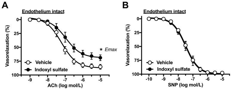
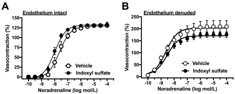
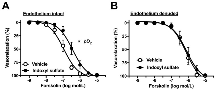
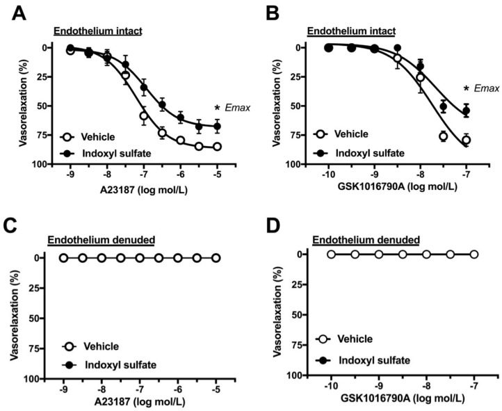
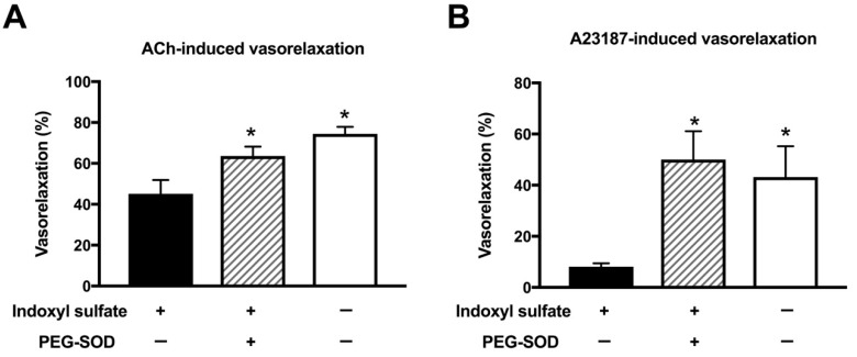
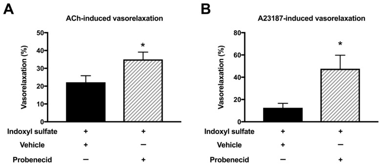
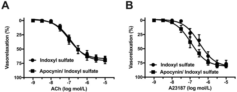
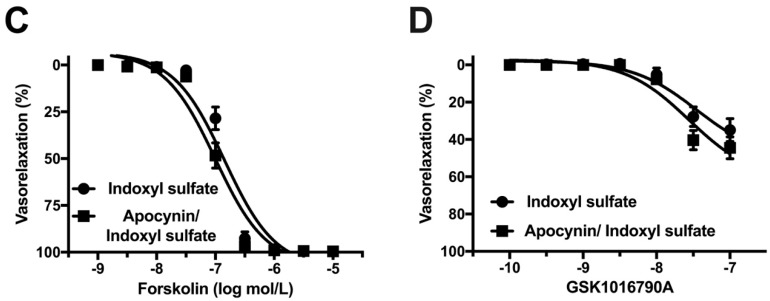
Gut microbiota are emerging as potential contributors to the regulation of host homeostasis. Dysbiosis of the gut microbiota associated with increased intestinal permeability facilitates the passage of endotoxins and other microbial products, including indoxyl sulfate in the circulation. Although an emerging body of evidence has suggested that indoxyl sulfate is a key substance for the development of chronic kidney disease, few studies have investigated the direct association of indoxyl sulfate with vascular function. We hypothesized that indoxyl sulfate adversely affects vascular function. Aortas isolated from male Wistar rat were examined in the presence or absence of indoxyl sulfate to assess the vascular function, including vasorelaxation and vasocontraction. Indoxyl sulfate (vs. vehicle) (1) decreased vasorelaxation induced by acetylcholine (ACh) but not by sodium nitroprusside; (2) had no significant alterations of noradrenaline-induced vasocontraction in the absence and presence of endothelium; (3) decreased adenylyl cyclase activator (forskolin)-induced vasorelaxation, while such a difference was eliminated by endothelial denudation; and (4) decreased vasorelaxations induced by calcium ionophore (A23187) and transient receptor potential vanilloid 4 agonist (GSK1016790A). The indoxyl sulfate-induced decrease in the vasorelaxations induced by ACh and A23187 increased by cell-permeant superoxide dismutase or by organic anion transporter inhibitor. However, apocynin, an inhibitor of nicotinamide adenine dinucleotide phosphate (NADPH) oxidase, had no effects on vasorelaxations induced by ACh, A23187, forskolin, and GSK1016790A in the presence of indoxyl sulfate. These results suggest that indoxyl sulfate directly affects the vascular function, particularly, endothelium-dependent vasorelaxation, and this effect may be attributable to increased oxidative stress after cell transportion via organic anion transporter, and such increased oxidative stress may not be attributable to activation of NADPH oxidase activation.

Keywords: Keywords: aorta; endothelial function; indoxyl sulfate; superoxide dismutase.


Conflict of interest statement

The authors declare that there are no conflict of interest.

Figures

Figure 1
Figure 1
Concentration–response curves for acetylcholine (ACh) (A) or sodium nitroprusside (SNP) (B)-induced vasorelaxations in the aortas in the absence (vehicle) and presence of indoxyl sulfate (10−4 mol/L). Ordinate shows vasorelaxation as a percentage of PE-induced vasocontraction (0% being defined as the plateau level of precontraction). Data are presented as mean ± standard error of mean (SEM) values from ten (A) or five (B) experiments. * p < 0.05, vs. vehicle; Emax.
Figure 2
Figure 2
Concentration–response curves for noradrenaline-induced vasocontractions in endothelium-intact (A) or -denuded (B) aortas in the absence (vehicle) and presence of indoxyl sulfate (10−4 mol/L). The ordinate shows vasocontraction as a percentage of 80 mmol/L high-K+-induced vasocontraction. Data are presented as means ± standard error of mean (SEM) values from six experiments.
Figure 3
Figure 3
Concentration–response curves for high K+-induced vasocontractions in the aortas in the absence (vehicle) and presence of indoxyl sulfate (10−4 mol/L). The ordinate shows vasocontraction as a percentage of 80 mmol/L high-K+-induced vasocontraction. Data are presented as mean ± standard error of mean (SEM) values from eight experiments.
Figure 4
Figure 4
Concentration–response curves for forskolin-induced vasorelaxations in endothelium-intact (A) or -denuded (B) aortas in the absence (vehicle) and presence of indoxyl sulfate (10−4 mol/L). The ordinate shows vasorelaxation as a percentage of PE-induced vasocontraction (0% being defined as the plateau level of precontraction). Data are presented as mean ± standard error of mean (SEM) values seven (A) or eight (B) experiments. * p < 0.05, vs. vehicle; a negative logarithm of EC50, which is the molar concentration of agonist producing 50% of the Emax (pD2).
Figure 5
Figure 5
Concentration–response curves for A23187 (A) or GSK1016790A (B)-induced vasorelaxations in endothelium-intact (A,C) or -denuded (B,D) aortas in the absence (vehicle) and presence of indoxyl sulfate (10−4 mol/L). The ordinate shows vasorelaxation as a percentage of PE-induced vasocontraction (0% being defined as the plateau level of precontraction). Data are presented as mean ± standard error of mean (SEM) values from eight (A) or six (BD) experiments. * p < 0.05, vs. Vehicle; Emax.
Figure 6
Figure 6
Effect of cell-permeant SOD on vasorelaxations induced by 3 × 10⁻⁷ mol/L acetylcholine (ACh) (A) or 10−7 mol/L A23187 (B) in the aorta treated with or without indoxyl sulfate. Polyethylene glycol-conjugated superoxide dismutase (PEG-SOD) (41 U/mL) was applied for 15 min; thereafter, indoxyl sulfate (10−4 mol/L) was incubated for 30 min before phenylephrine (PE) application. The ordinate shows vasorelaxation as a percentage of PE-induced vasocontraction (0% being defined as the plateau level of precontraction). Data are presented as mean ± standard error of mean (SEM) values from six experiments. * p < 0.05 vs. indoxyl sulfate group.
Figure 7
Figure 7
Effect of organic acid transporter inhibitor on vasorelaxations induced by 3 × 10⁻⁷ mol/L acetylcholine (ACh) (A) or 10−7 mol/L A23187 (B) in the aorta treated with indoxyl sulfate. Probenecid (10−3 mol/L) or vehicle (ethanol) was applied for 30 min; thereafter, indoxyl sulfate (10−4 mol/L) was incubated for 30 min before phenylephrine (PE) application. The ordinate shows vasorelaxation as a percentage of the PE-induced vasocontraction (0% being defined as the plateau level of precontraction). Data are presented as mean ± standard error of mean (SEM) values from six experiments. * p < 0.05 vs. indoxyl sulfate group.
Figure 8
Figure 8
Effect of an NADPH oxidase inhibitor on vasorelaxations induced by acetylcholine (ACh) (A), A23187 (B), forskolin (C), or GSK1016790A (D) in the aorta treated with indoxyl sulfate. Apocynin (10−4 mol/L) or vehicle (DMSO) was applied for 30 min; thereafter, indoxyl sulfate (10−4 mol/L) was incubated for 30 min before phenylephrine (PE) application. The ordinate shows vasorelaxation as a percentage of the PE-induced vasocontraction (0% being defined as the plateau level of precontraction). Data are presented as mean ± standard error of mean (SEM) values from eleven (A), ten (B), and twelve (C,D) experiments.
Figure 8
Figure 8
Effect of an NADPH oxidase inhibitor on vasorelaxations induced by acetylcholine (ACh) (A), A23187 (B), forskolin (C), or GSK1016790A (D) in the aorta treated with indoxyl sulfate. Apocynin (10−4 mol/L) or vehicle (DMSO) was applied for 30 min; thereafter, indoxyl sulfate (10−4 mol/L) was incubated for 30 min before phenylephrine (PE) application. The ordinate shows vasorelaxation as a percentage of the PE-induced vasocontraction (0% being defined as the plateau level of precontraction). Data are presented as mean ± standard error of mean (SEM) values from eleven (A), ten (B), and twelve (C,D) experiments.

Similar articles

Cited by

References

    1. Jie Z., Xia H., Zhong S.L., Feng Q., Li S., Liang S., Zhong H., Liu Z., Gao Y., Zhao H., et al. The gut microbiome in atherosclerotic cardiovascular disease. Nat. Commun. 2017;8:845. doi: 10.1038/s41467-017-00900-1. - DOI - PMC - PubMed
    1. Jonsson A.L., Backhed F. Role of gut microbiota in atherosclerosis. Nat. Rev. Cardiol. 2017;14:79–87. doi: 10.1038/nrcardio.2016.183. - DOI - PubMed
    1. Karlsson F., Tremaroli V., Nielsen J., Backhed F. Assessing the human gut microbiota in metabolic diseases. Diabetes. 2013;62:3341–3349. doi: 10.2337/db13-0844. - DOI - PMC - PubMed
    1. Koren O., Spor A., Felin J., Fak F., Stombaugh J., Tremaroli V., Behre C.J., Knight R., Fagerberg B., Ley R.E., et al. Human oral, gut, and plaque microbiota in patients with atherosclerosis. Proc. Natl. Acad. Sci. USA. 2011;108:4592–4598. doi: 10.1073/pnas.1011383107. - DOI - PMC - PubMed
    1. Lau W.L., Savoj J., Nakata M.B., Vaziri N.D. Altered microbiome in chronic kidney disease: Systemic effects of gut-derived uremic toxins. Clin. Sci. 2018;132:509–522. doi: 10.1042/CS20171107. - DOI - PubMed

MeSH terms

LinkOut - more resources