. 2019 Jan 15;11(1):97.
doi: 10.3390/cancers11010097.

On the Mechanism of Hyperthermia-Induced BRCA2 Protein Degradation

Affiliations

On the Mechanism of Hyperthermia-Induced BRCA2 Protein Degradation

Nathalie van den Tempel et al. Cancers (Basel). .

Abstract

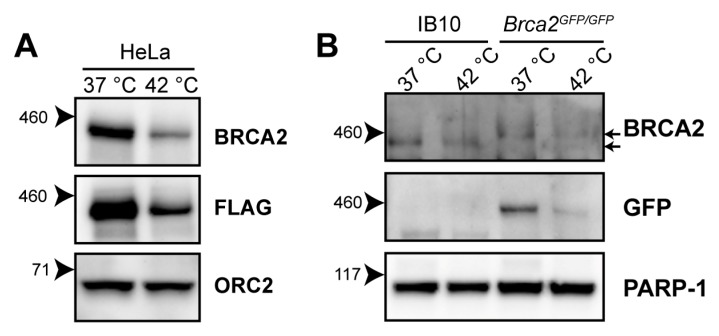
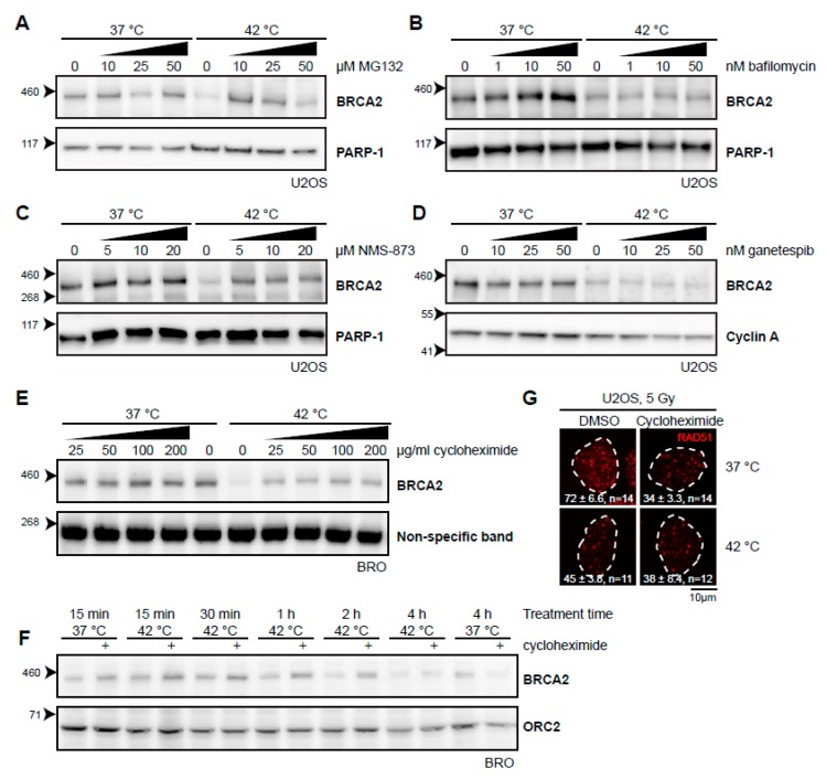
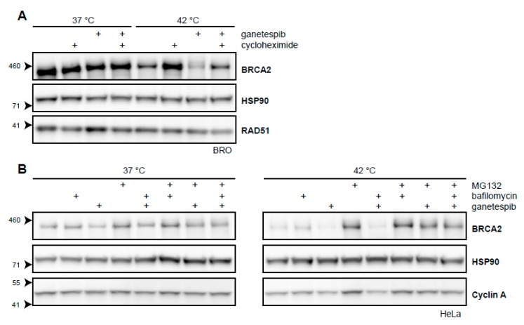
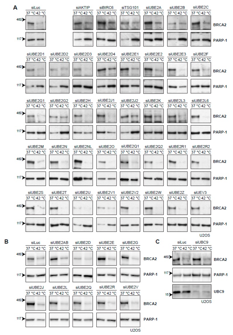
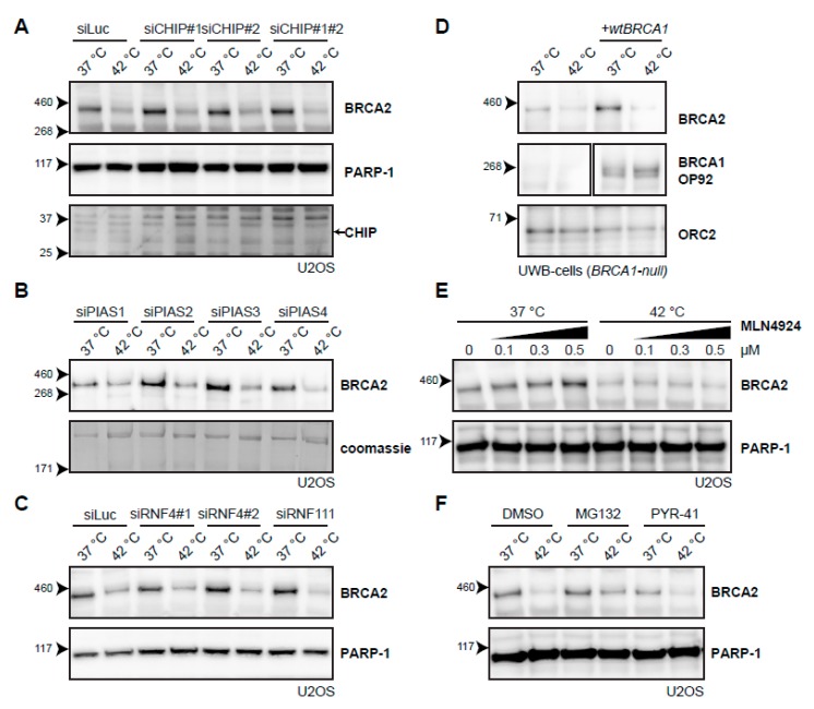
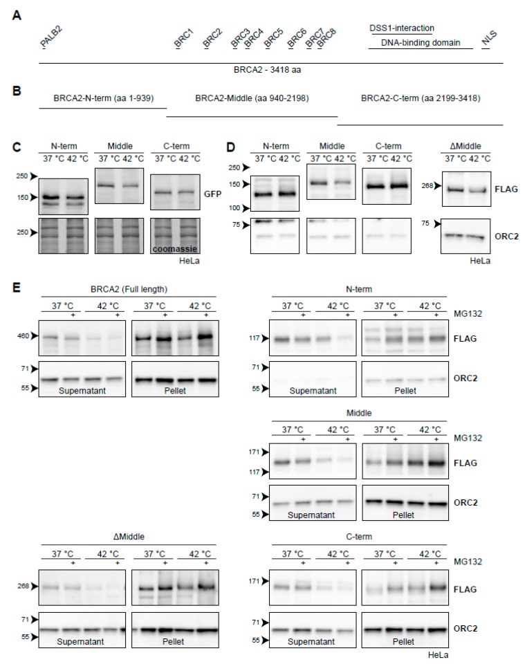
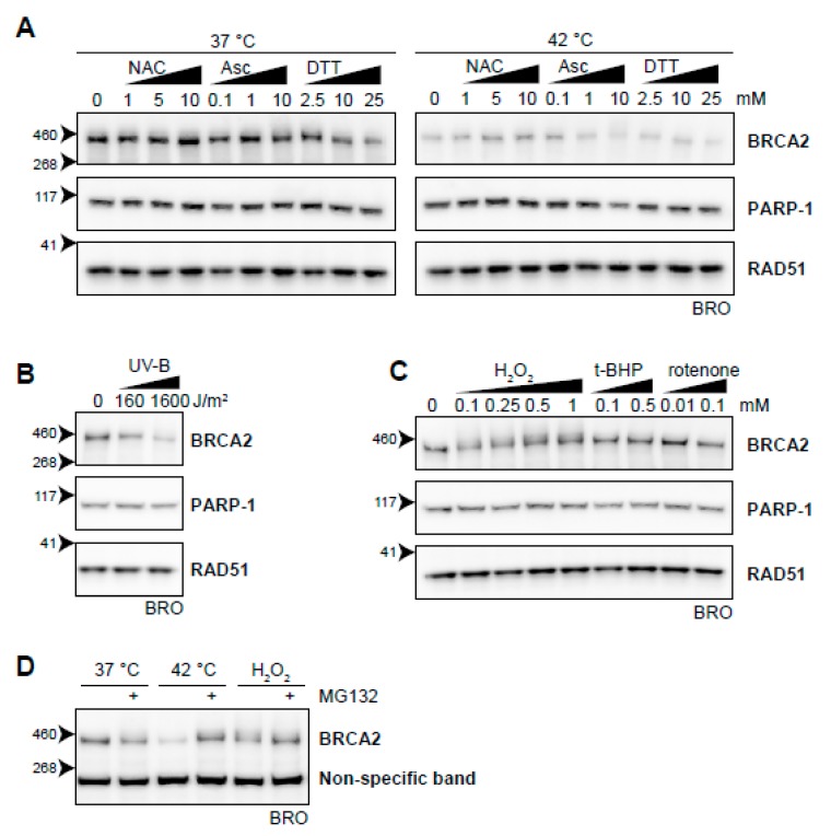
The DNA damage response (DDR) is a designation for a number of pathways that protects our DNA from various damaging agents. In normal cells, the DDR is extremely important for maintaining genome integrity, but in cancer cells these mechanisms counteract therapy-induced DNA damage. Inhibition of the DDR could therefore be used to increase the efficacy of anti-cancer treatments. Hyperthermia is an example of such a treatment-it inhibits a sub-pathway of the DDR, called homologous recombination (HR). It does so by inducing proteasomal degradation of BRCA2 -one of the key HR factors. Understanding the precise mechanism that mediates this degradation is important for our understanding of how hyperthermia affects therapy and how homologous recombination and BRCA2 itself function. In addition, mechanistic insight into the process of hyperthermia-induced BRCA2 degradation can yield new therapeutic strategies to enhance the effects of local hyperthermia or to inhibit HR. Here, we investigate the mechanisms driving hyperthermia-induced BRCA2 degradation. We find that BRCA2 degradation is evolutionarily conserved, that BRCA2 stability is dependent on HSP90, that ubiquitin might not be involved in directly targeting BRCA2 for protein degradation via the proteasome, and that BRCA2 degradation might be modulated by oxidative stress and radical scavengers.

Keywords: BRCA2; HSP90; RAD51; SILAC mass spectrometry; homologous recombination; hyperthermia; protein degradation; reactive oxygen species; ubiquitin.


Conflict of interest statement

The authors declare no conflict of interest

Figures

Figure 1
Figure 1
Heat-mediated degradation of BRCA2 and modulation of homologous recombination (HR). (A) Immunoblot of HeLa cells stably expressing expressing BRCA2, tagged with the FLAG epitope (DYKDDDDK), treated with or without 60 min of hyperthermia. (B) Immunoblot of wild-type IB10 and BRCA2GFP/GFP mouse embryonic stem (mES) cells. The upper and lower arrow next to the upper BRCA2 panel indicate the positions of BRCA2-GFP and untagged BRCA2, respectively.
Figure 2
Figure 2
Various inhibitors alter the heat-mediated BRCA2 degradation. (AE) Immunoblots of cells treated with or without 60 min of hyperthermia in the presence of indicated doses of different inhibitors. All inhibitors were added 30–60 min prior to hyperthermia treatment. (A) Human Bone Osteosarcoma U2OS cells treated with the proteasome inhibitor MG132. (B) HeLa cells treated with the autophagy inhibitor bafilomycin A1. (C) U2OS cells treated with an inhibitor of the valosin-containing protein (VCP) segregase. (D) U2OS cells treated with the HSP90 inhibitor, ganetespib. (E) Lymphoma-derived BRO cells treated with the translation inhibitor, cycloheximide. (F) BRO cells treated with cycloheximide (50 µg/mL) and hyperthermia for the indicated periods of time. (G) U2OS cells were treated with or without hyperthermia in the presence of dimethylsulfoxide (DMSO) and cycloheximide, irradiated with 5 Gy, and fixed 90 min after irradiation. Cells were stained for 5-ethynyl-2’-deoxyuridine (EdU) and the protein product of the Radiation 51 RAD51 gene. The panel shows the RAD51-staining for representative cells for each condition. The dotted line indicates the perimeter of an EdU-positive nucleus. Numbers in the panel indicate the mean number of foci ± standard error of the mean and the number of cells analyzed.
Figure 3
Figure 3
BRCA2 is a client protein of HSP90. (A) Immunoblot of BRO cells treated with or without 60 min of hyperthermia in the presence of cycloheximide (50 µg/mL), ganetespib (50 nM), or both. (B) Immunoblots of HeLa cells treated with or without hyperthermia in the presence of various combinations of proteasomal inhibitor MG132 (50 µM), autophagy inhibitor bafilomycin (10 nM), and HSP90-inhibitor ganetespib (50 nM).
Figure 4
Figure 4
Efforts to identify the E2-conjugating enzyme that mediates degradation of BRCA2 upon hyperthermia. (A) Results of individual knock-down of all known ubiquitin E2-conjugating enzymes in U2OS cells. Efficiencies of E2 siRNAs were previously assessed by quantitative polymerase chain reaction PCR [37]. (B) Results of simultaneous knock-downs of E2-conjugating enzymes belonging to specific families in U2OS cells. (C) Results of knock-down of the small ubiquitin-like modifier (SUMO)-E2 enzyme Ubiquitin Conjugating Enzyme 9 (UBC9) in U2OS cells.
Figure 5
Figure 5
Searching for the E3-ligase that mediates degradation of BRCA2 upon hyperthermia. (A) Effect of hyperthermia on BRCA2 levels upon knock-down of the Heat-shock Protein (HSP)-associated E3-ligase, C terminus of HSC70-Interacting Protein (CHIP), in U2OS cells using two different siRNAs. Arrow on the right next to the CHIP panel indicates the predicted position of the CHIP protein. (B) Effect of hyperthermia on BRCA2 levels upon knock-down of the SUMO-E3 ligases Protein Inhibitor Of Activated STAT( PIAS) 1-4 in U2OS cells using siRNAs. (C) Effect of hyperthermia on BRCA2 levels upon siRNA knock-down of DNA-repair-related, SUMO-targeted ubiquitin ligases, Ring Finger (RNF) 4 and RNF111. (D) Detection of BRCA2 and BRCA1 in UWB1.289 (BRCA1-null) cells with or without complementation of wild type BRCA1 (wtBRCA1). (E) Detection of BRCA2 in U2OS cells treated with increased doses of the neddylation-inhibitor MLN4924, added 30 min prior to hyperthermia. (F) Detection of BRCA2 in U2OS cells treated with or without hyperthermia in the presence of proteasome inhibitor MG132 (10 µM) or the E1-activating enzyme inhibitor PYR-41 (10 µM). Inhibitors were added 1 h prior to hyperthermia treatment, and left on for the duration of the treatment.
Figure 6
Figure 6
BRCA2 degradation upon heat treatment from the perspective of BRCA2. (A) Graphical representation of the full-length 3418-aa BRCA2-protein. Interaction domains are indicated as small bars above the full-length protein. (B) Arbitrary truncations of the BRCA2-protein to be tested for heat stability. (C) HeLa cells stably expressing GFP-BRCA2 fragments. (D) HeLa cells stably expressing FLAG-BRCA2 fragments. The right panel contains a FLAG-tagged ΔMiddle construct, which effectively is a fusion of the constructs designated as the N and C-termini. (E) Hela cells expressing the different constructs were subjected to hyperthermia in the absence or presence of 50 µM MG132 and processed following a fractionation protocol prior to lysis. The resulting supernatant and pellet fractions are shown.
Figure 7
Figure 7
Oxidative stress as an inducer of BRCA2 degradation. (A) BRCA2 levels in BRO cells subjected to hyperthermia in the presence of indicated doses of antioxidant compounds N-acetylcysteine (NAC), ascorbic Acid (Asc), or dithiothreitol (DTT). (B) BRCA2 levels in BRO cells subjected to the indicated dose of ultraviolet-B and lysed three hours afterwards. (C) BRCA2 levels in BRO cells treated with various concentrations of oxidative stress-inducers H2O2, tert-Butyl hydroperoxide (t-BHP), and Rotenone. (D) BRCA2 levels in BRO cells exposed to hyperthermia or H2O2 (0.1 mM) in the presence or absence of proteasome inhibitor MG132 (50 µM).

References

    1. Hoeijmakers J.H. Genome maintenance mechanisms for preventing cancer. Nature. 2001;411:366–374. doi: 10.1038/35077232. - DOI - PubMed
    1. Jackson S.P., Bartek J. The DNA-damage response in human biology and disease. Nature. 2009;461:1071–1078. doi: 10.1038/nature08467. - DOI - PMC - PubMed
    1. Ciccia A., Elledge S.J. The DNA damage response: Making it safe to play with knives. Mol. Cell. 2010;40:179–204. doi: 10.1016/j.molcel.2010.09.019. - DOI - PMC - PubMed
    1. Tian H., Gao Z., Li H., Zhang B., Wang G., Zhang Q., Pei D., Zheng J. DNA damage response—A double-edged sword in cancer prevention and cancer therapy. Cancer Lett. 2015;358:8–16. doi: 10.1016/j.canlet.2014.12.038. - DOI - PubMed
    1. Curtin N.J. Inhibiting the DNA damage response as a therapeutic manoeuvre in cancer. Br. J. Pharmacol. 2013;169:1745–1765. doi: 10.1111/bph.12244. - DOI - PMC - PubMed