. 2019 Mar;7(3):363-375.
doi: 10.1158/2326-6066.CIR-18-0572. Epub 2019 Jan 16.

NK Cells Expressing a Chimeric Activating Receptor Eliminate MDSCs and Rescue Impaired CAR-T Cell Activity against Solid Tumors

Affiliations

NK Cells Expressing a Chimeric Activating Receptor Eliminate MDSCs and Rescue Impaired CAR-T Cell Activity against Solid Tumors

Robin Parihar et al. Cancer Immunol Res. 2019 Mar.

Abstract

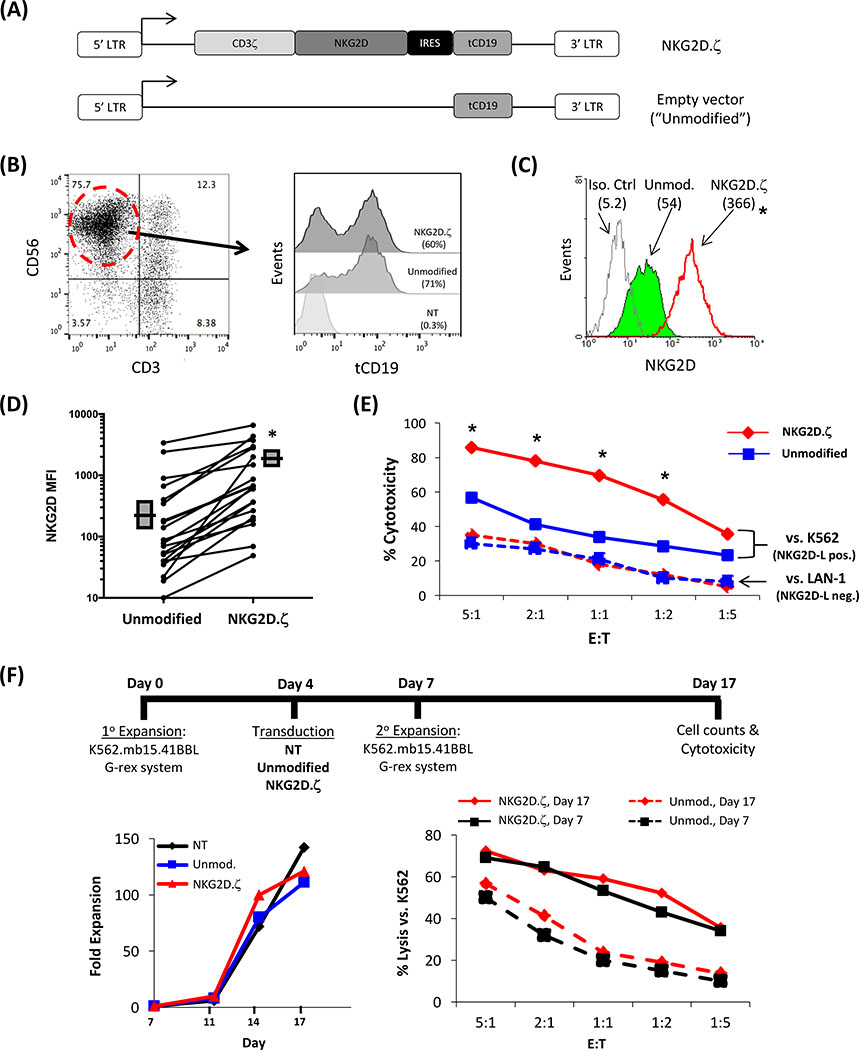
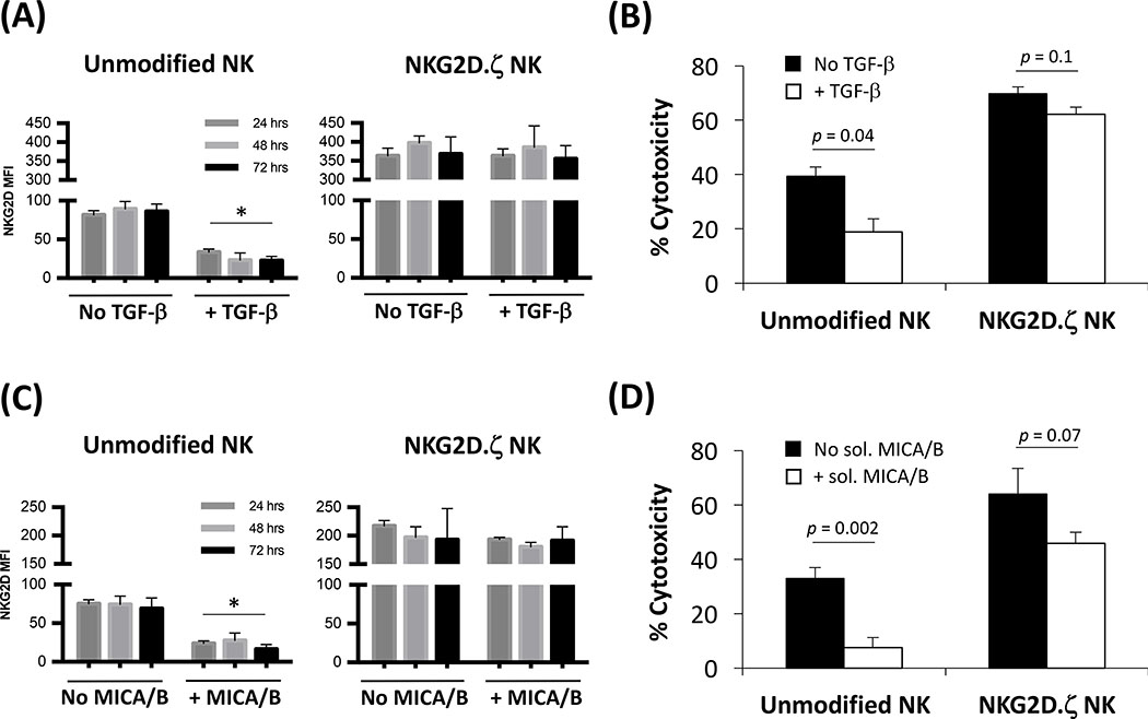
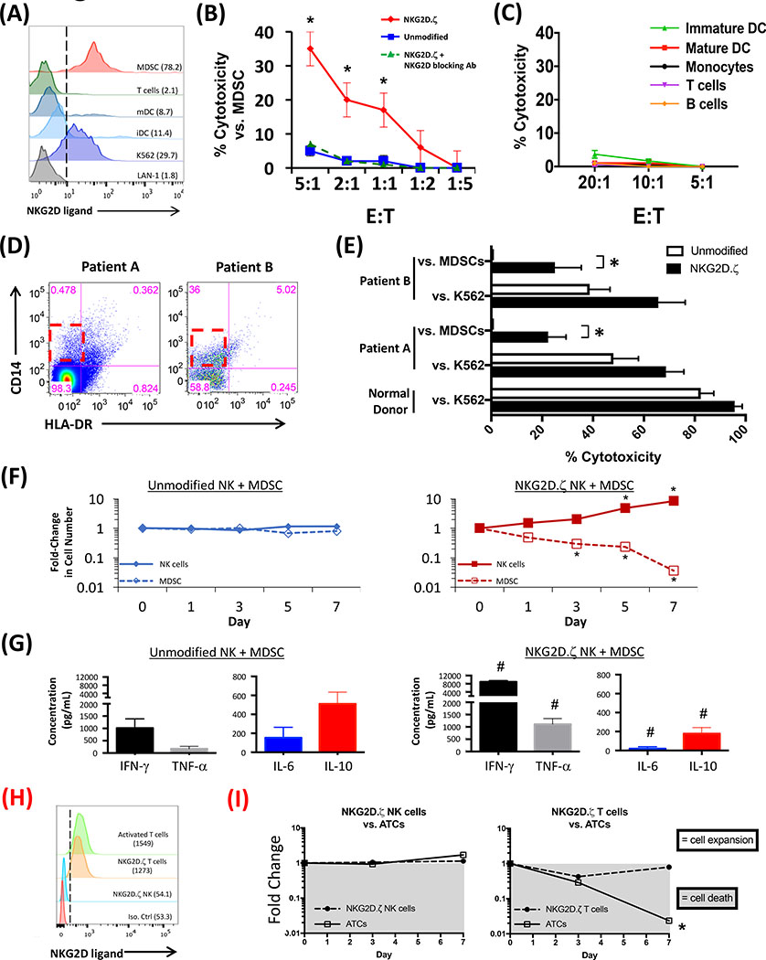
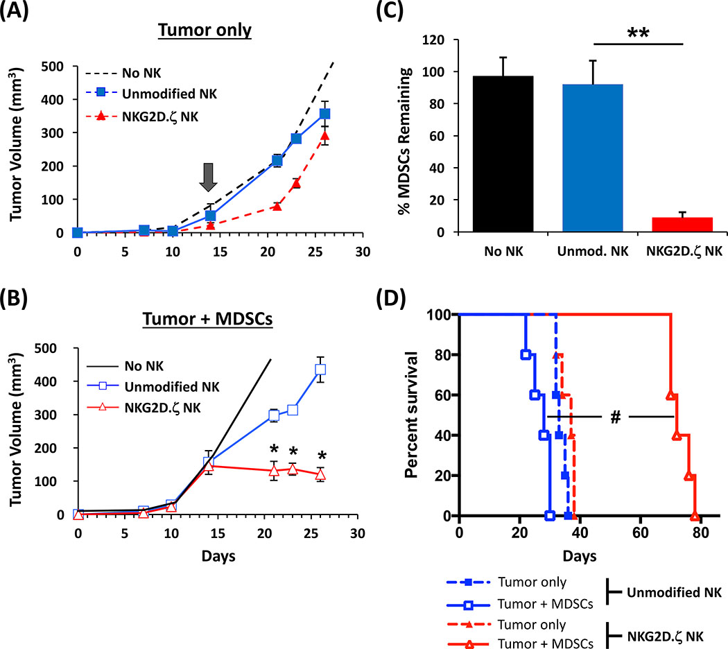
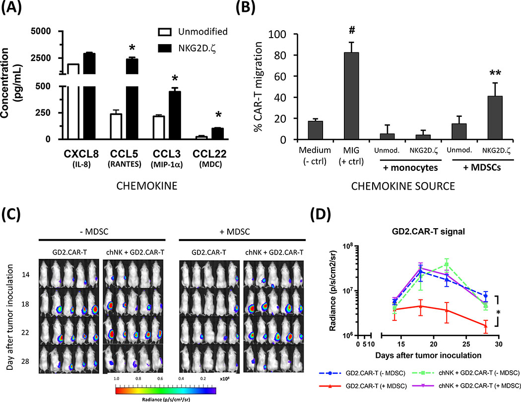
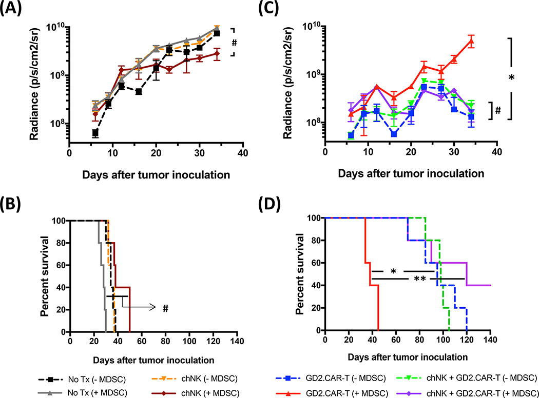
Solid tumors are refractory to cellular immunotherapies in part because they contain suppressive immune effectors such as myeloid-derived suppressor cells (MDSCs) that inhibit cytotoxic lymphocytes. Strategies to reverse the suppressive tumor microenvironment (TME) should also attract and activate immune effectors with antitumor activity. To address this need, we developed gene-modified natural killer (NK) cells bearing a chimeric receptor in which the activating receptor NKG2D is fused to the cytotoxic ζ-chain of the T-cell receptor (NKG2D.ζ). NKG2D.ζ-NK cells target MDSCs, which overexpress NKG2D ligands within the TME. We examined the ability of NKG2D.ζ-NK cells to eliminate MDSCs in a xenograft TME model and improve the antitumor function of tumor-directed chimeric antigen receptor (CAR)-modified T cells. We show that NKG2D.ζ-NK cells are cytotoxic against MDSCs, but spare NKG2D ligand-expressing normal tissues. NKG2D.ζ-NK cells, but not unmodified NK cells, secrete proinflammatory cytokines and chemokines in response to MDSCs at the tumor site and improve infiltration and antitumor activity of subsequently infused CAR-T cells, even in tumors for which an immunosuppressive TME is an impediment to treatment. Unlike endogenous NKG2D, NKG2D.ζ is not susceptible to TME-mediated downmodulation and thus maintains its function even within suppressive microenvironments. As clinical confirmation, NKG2D.ζ-NK cells generated from patients with neuroblastoma killed autologous intratumoral MDSCs capable of suppressing CAR-T function. A combination therapy for solid tumors that includes both NKG2D.ζ-NK cells and CAR-T cells may improve responses over therapies based on CAR-T cells alone.


Conflict of interest statement

The authors declare no potential conflicts of interest.

Figures

Figure 1.
Figure 1.. NKG2D.ζ–NK cells expand and kill ligand-expressing targets.
(A) Schematic of SFG-based retroviral vector constructs for transduction of human NK cells. (B) Human NK cells were expanded as described in Methods and percentage of CD56+/CD3 NK cells at time of retroviral transduction (day 4) is shown. Expanded NK cells (red circle in A) purified via depletion of CD3+ cells were transduced with NKG2D.ζ retroviral vector or empty vector control (referred to as “unmodified”), and transduction efficiencies are shown inset. (C) NKG2D expression on NK cells (MFI, inset) was assessed with isotype antibody as control. Non-transduced NK cells exhibited similar NKG2D expression to empty vector-transduced NK cells. * p = 0.003 vs. unmodified condition. (D) Expression of NKG2D (absolute MFI on y-axis) on NK cells from each donor (n = 25) transduced with either empty vector or NKG2D.ζ construct was determined by flow cytometry. Each pair of data points connected by a line represent cells from a single donor, to confirm surface expression of our chimeric molecule after transduction. Black line with grey block next to each group are mean MFI ± SEM. (E) NKG2D.ζ–NK cell cytotoxicity against K562 and LAN-1 tumor targets in a 4-hour 51Cr-release assay. Given that K562 and LAN-1 are both NK-sensitive targets, low E:T ratios were utilized to observe differences. Experiment is representative of at least three separate determinations from n = 10 donors. * p < 0.01 vs. unmodified NK cells at same E:T ratio. (F) NKG2D.ζ–NK cells were expanded after transduction culture (as shown in schema), and fold-expansion and cytotoxicity both pre- (day 7) and post- (day 17) secondary expansion were determined.
Figure 2.
Figure 2.. Transgenic NKG2D.ζ is unaffected by TGFβ or soluble NKG2D ligands.
NKG2D.ζ or unmodified NK cells (n = 5 donors) were cultured in the presence of TGFβ (5 ng/mL) (A, B) or the soluble NKG2D ligands MICA and MICB (C, D) for 24-, 48-, and 72-hours. NKG2D receptor expression was determined by flow cytometry and NK cytotoxicity against K562 targets was assessed in a 4-hr Cr-release assay at an 5:1 E:T ratio using 48-hr exposed NK cells. Viability of transduced NK cells after exposure to TGFβ for 24, 48, and 72 hours, as assessed by 7-AAD vital staining, was > 90%. * p = 0.001 vs. non-TGFβ/MICA-treated NK groups at same time-points.
Figure 3.
Figure 3.. Human MDSCs express ligands for NKG2D and are killed by NKG2D.ζ–NK cells.
(A) NKG2D ligand expression on human MDSCs by flow cytometry. Immature dendritic cells (iDC) and mature DCs (mDC) were used as myeloid controls. T cells activated with CD3 and CD28 mAbs plus 100 IU/mL IL2 for 24 hours were used as lymphocyte control. LAN-1 and K562 cells were used as negative and positive controls, respectively. MFI of NKG2D ligand expression in parenthesis. Representative data from single donor (of n = 25 normal donors). Isotype control for NKG2D staining routinely fell within the 1st log. (B) NKG2D.ζ–NK cell cytotoxicity against autologous MDSCs as targets in a 4-hour 51Cr-release assay. In some wells of the cytotoxicity assay, a blocking mAb to NKG2D was added. Representative data from triplicate samples per data point from a single donor (of n = 25 normal donors) is shown. * p < 0.01 vs. unmodified NK cells at same E:T ratio. (C) In the same experiment as (B), the same batch of NKG2D.ζ–NK cells were analyzed for cytotoxicity against autologous B cells, monocytes, monocyte-derived iDC and mDC, and activated T cells (n = 10 donors examined). (D) M-MDSC frequency by flow cytometry from neuroblastoma tumor samples obtained from high-risk patients, as described in Methods. (E) Cytotoxicity by NKG2D.ζ–NK cells derived from patient PBMC (harvested and frozen at time of tumor sampling) against autologous tumor-derived MDSCs in a 4-hour 51Cr-release assay. Data shown are from triplicate samples per data point at a 10:1 E:T ratio. * p < 0.001 vs. unmodified NK cells from same donor. (F) NKG2D.ζ–NK cells were cocultured with autologous MDSCs at 1:1 ratio plus low-dose 50 IU/mL IL2 to maintain NK survival, and fold change in the number of each cell type from the start of coculture was determined by flow cytometry at indicated time-points. * p < 0.001 vs. NK/MDSC fold-change in unmodified NK cell cocultures. (G) Cell-free supernatants were harvested from cocultures at day 3 and analyzed for IFNγ, TNFα, IL6, and IL10 by ELISA. # p < 0.01 vs. corresponding cytokine in cocultures with unmodified NK cells. (H) NKG2D ligand expression was determined for activated T cells (ATCs) expressing NKG2D.ζ and NKG2D.ζ–NK cells. Expression of NKG2D ligands on non-transduced ATCs as control for T-cell activation. (I) NKG2D.ζ–NK cells or NKG2D.ζ T cells were cocultured with autologous ATCs at 1:1 ratio and fold change in the number of each cell type from the start of coculture was determined by flow cytometry at indicated time-points. * p < 0.001 vs. ATC fold-change at days 0 and 3 cocultures.
Figure 4.
Figure 4.. NKG2D.ζ–NK cells eliminate intra-tumoral MDSCs and reduce tumor burden.
LAN-1 tumor cells, either alone (A) or admixed with human MDSCs (B), were injected S.C. in the flanks of NSG mice. When tumors reached a volume of approx. 100 mm3 (day 14, gray block arrow inset), no NK cells (PBS control), 1×10⁷ unmodified or NKG2D.ζ–NK cells were injected I.V. and tumor growth was measured over time via calipers. * p < 0.03 vs. other conditions shown at same time point. (C) On day 26, intra-tumoral human MDSCs (CD33+, HLA-DRlow) were enumerated by flow cytometry and are presented as mean % MDSCs remaining per treatment group. ** p < 0.005 vs. unmodified NK treatment. (D) Survival of groups by Kaplan-Meyer analysis. # p = 0.024. Representative experiment of three performed.
Figure 5.
Figure 5.. NKG2D.ζ–NK cells secrete chemokines that recruit GD2.CAR-T cells.
(A) NKG2D.ζ or unmodified NK cells were cocultured with autologous MDSCs and cell-free culture supernatants harvested at 48 hours were analyzed for chemokines CXCL8, CCL5, CCL3, and CCL22 by ELISA. Shown are mean chemokine concentration ± SEM for n = 5 cocultures/donor (data from one of five representative donors is shown). * p < 0.005 vs. unmodified NK cocultures. (B) GD2.CAR-T cells were assayed for chemotaxis in Transwells (described in Methods) in response to supernatants derived from unmodified or NKG2D.ζ–NK cells cocultured with autologous MDSCs. Supernatants derived from monocyte (non-suppressive myeloid cell control)-stimulated NK cells were also used. # p < 0.001 vs. Medium, ** p = 0.009 vs. “unmodified NK plus MDSCs” condition. (C) LAN-1 tumor cells, alone or admixed with human MDSCs, were injected S.C. into the flank of NSG mice. When tumors reached a volume ~100 mm3, 5×10⁶ GD2.CAR-T cells were injected I.V. alone on day 13 (GD2.CAR-T), or preceded by 5×10⁶ NKG2D.ζ–NK cells I.V. injected on day 10 (chNK + GD2.CAR-T). GD2.CAR-T signal at tumor site was measured over time via live-animal bioluminescence imaging. (D) Shown is mean ± SEM (n=5 mice/group) bioluminescent signal of GD2.CAR-T cells expressed as radiance. * p = 0.01 vs. all other groups.
Figure 6.
Figure 6.. Elimination of MDSCs by NKG2D.ζ–NK cells increases antitumor activity of GD2.CAR-T cells.
Luciferase gene-transduced LAN-1 tumor cells, alone or admixed with human MDSCs, were injected S.C. into NSG mice. (A) When tumors reached a volume ~100 mm3, no treatment (No Tx; PBS control) or 5×10⁶ NKG2D.ζ–NK cells alone (chNK) were injected I.V. on day 10 and tumor growth was measured over time via live-animal bioluminescence imaging. Shown is mean ± SEM (n=5 mice/group) bioluminescent signal expressed as radiance. # ns, p = 0.18 vs. No treatment (+MDSC) group. (B) Survival of groups in A was determined by Kaplan-Meyer analysis. # ns, p = 0.059. (C) In other groups of mice within the same experiment, 5×10⁶ GD2.CAR-T cells were injected I.V. alone on day 13 (GD2.CAR-T), or preceded by 5×10⁶ NKG2D.ζ–NK cells injected on day 10 (chNK+GD2.CAR-T). * p = 0.001; # ns, p = 0.59 vs. each other. (D) Survival of groups in C by Kaplan-Meyer analysis. Representative experiment of 5 separate experiments. * p = 0.002; ** p = 0.001.

Similar articles

Cited by

References

    1. Fesnak AD, June CH, and Levine BL. Engineered T cells: the promise and challenges of cancer immunotherapy. Nat Rev Cancer. 2016;16(9):566–81. - PMC - PubMed
    1. Maude SL, Frey N, Shaw PA, Aplenc R, Barrett DM, Bunin NJ, Chew A, Gonzalez VE, Zheng Z, Lacey SF, et al. Chimeric antigen receptor T cells for sustained remissions in leukemia. N Engl J Med. 2014;371(16):1507–17. - PMC - PubMed
    1. Davila ML, Riviere I, Wang X, Bartido S, Park J, Curran K, Chung SS, Stefanski J, Borquez-Ojeda O, Olszewska M, et al. Efficacy and toxicity management of 19–28z CAR T cell therapy in B cell acute lymphoblastic leukemia. Sci Transl Med. 2014;6(224):224ra25. - PMC - PubMed
    1. Hege KM, Bergsland EK, Fisher GA, Nemunaitis JJ, Warren RS, McArthur JG, Lin AA, Schlom J, June CH, and Sherwin SA. Safety, tumor trafficking and immunogenicity of chimeric antigen receptor (CAR)-T cells specific for TAG-72 in colorectal cancer. J Immunother Cancer. 2017;5(22. - PMC - PubMed
    1. Thistlethwaite FC, Gilham DE, Guest RD, Rothwell DG, Pillai M, Burt DJ, Byatte AJ, Kirillova N, Valle JW, Sharma SK, et al. The clinical efficacy of first-generation carcinoembryonic antigen (CEACAM5)-specific CAR T cells is limited by poor persistence and transient pre-conditioning-dependent respiratory toxicity. Cancer immunology, immunotherapy : CII. 2017;66(11):1425–36. - PMC - PubMed

Publication types

MeSH terms