. 2019 Jan 16;11(475):eaat5580.
doi: 10.1126/scitranslmed.aat5580.

Clinical-grade stem cell-derived retinal pigment epithelium patch rescues retinal degeneration in rodents and pigs

Affiliations

Clinical-grade stem cell-derived retinal pigment epithelium patch rescues retinal degeneration in rodents and pigs

Ruchi Sharma et al. Sci Transl Med. .

Abstract

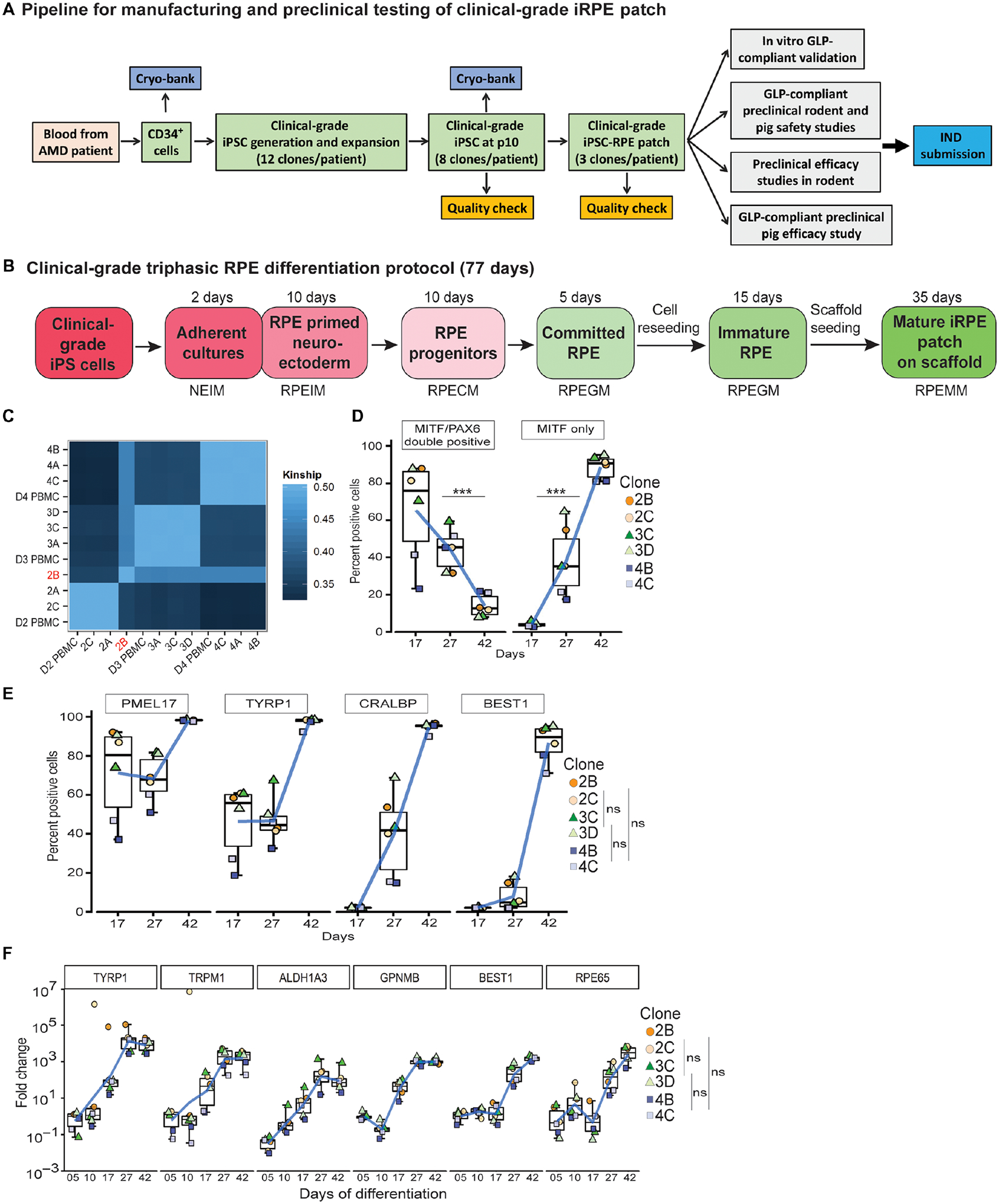
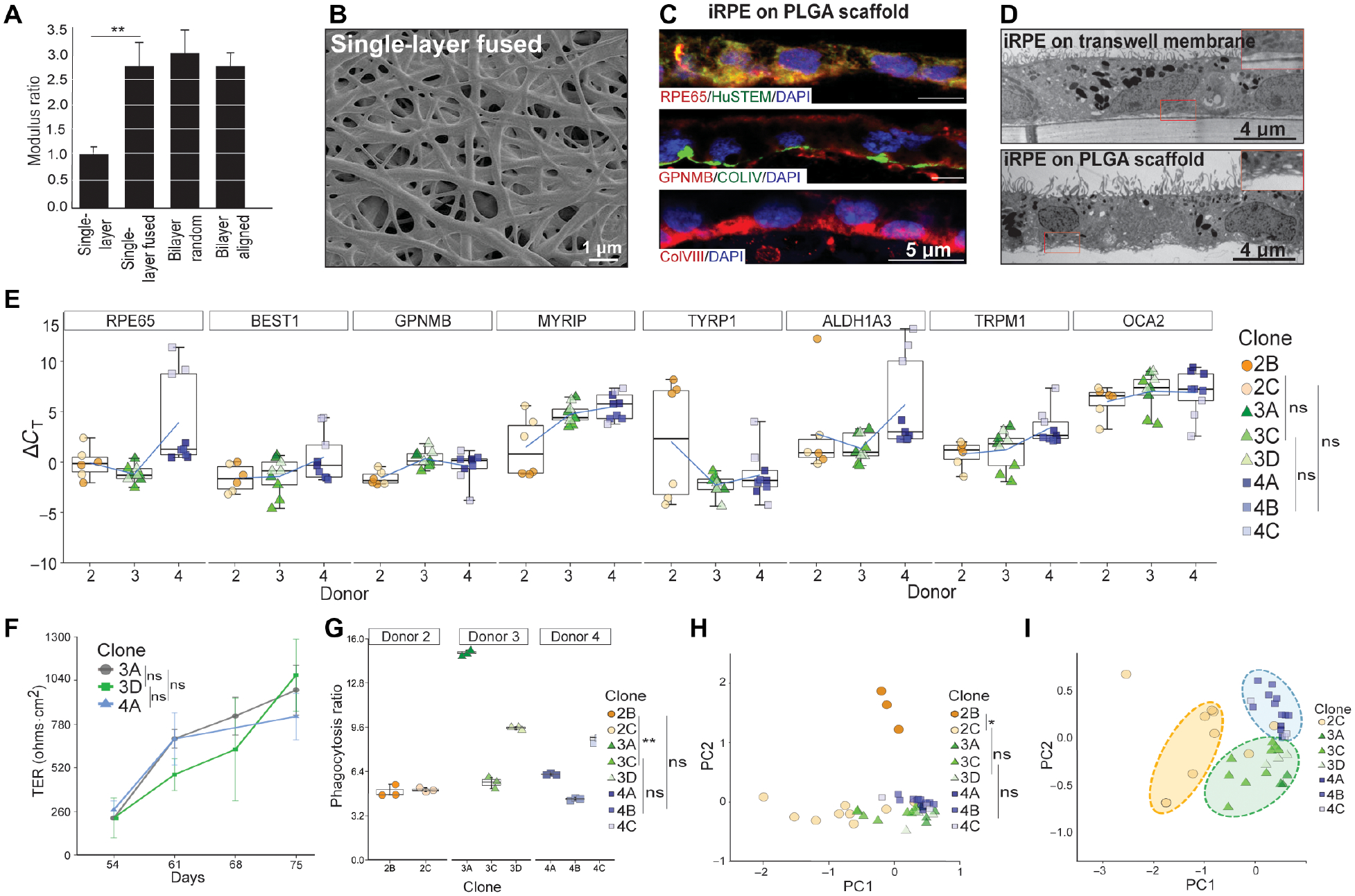
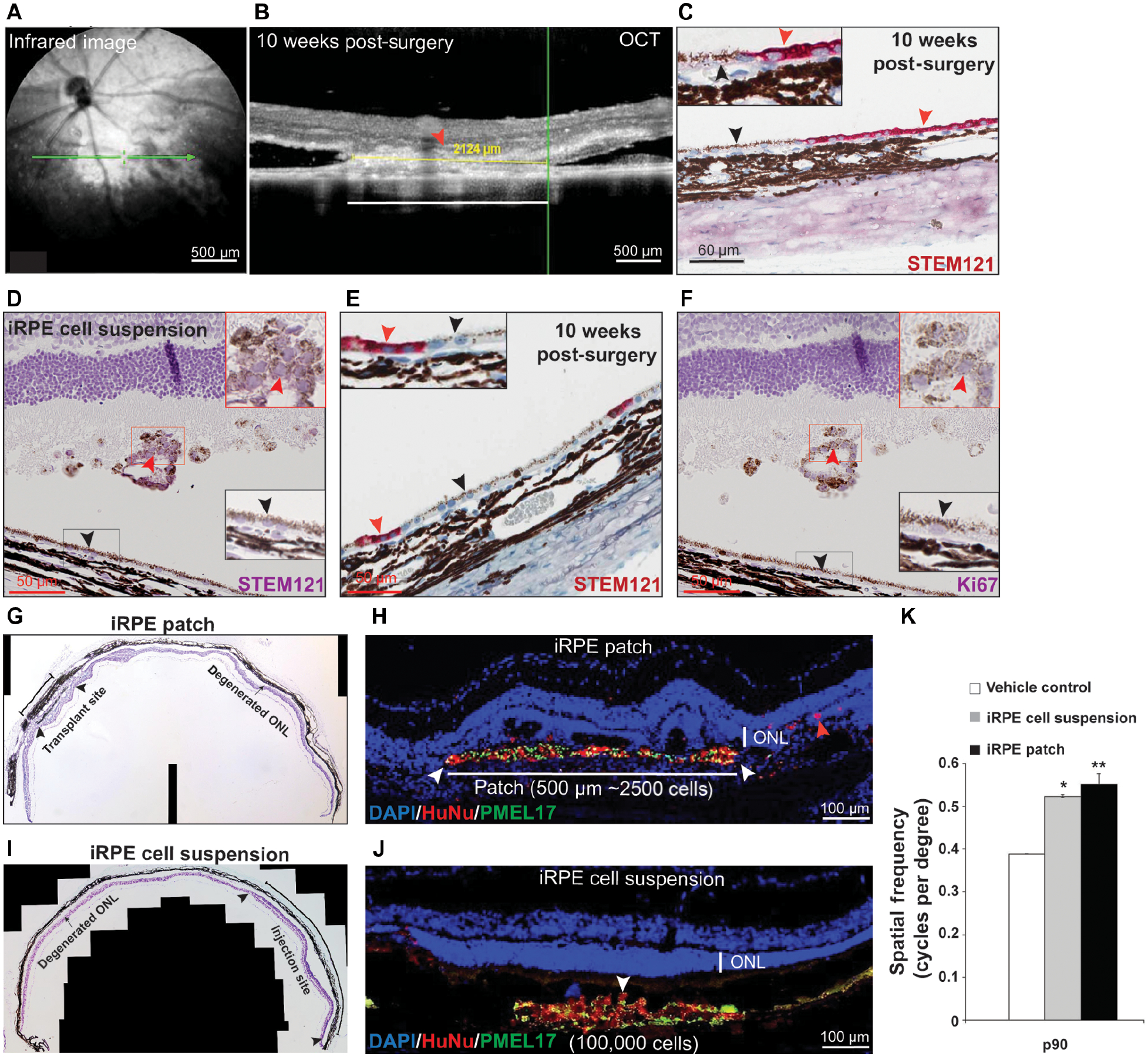
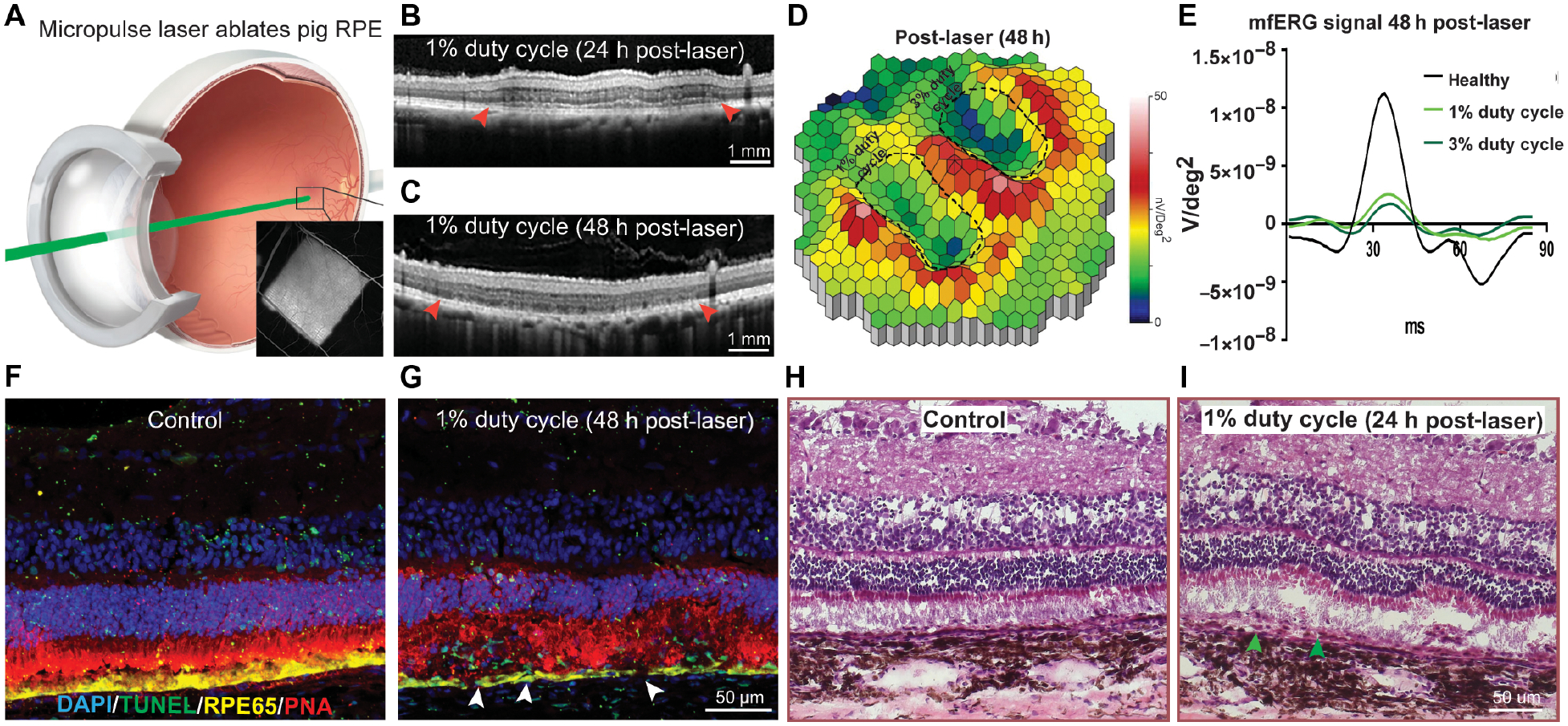
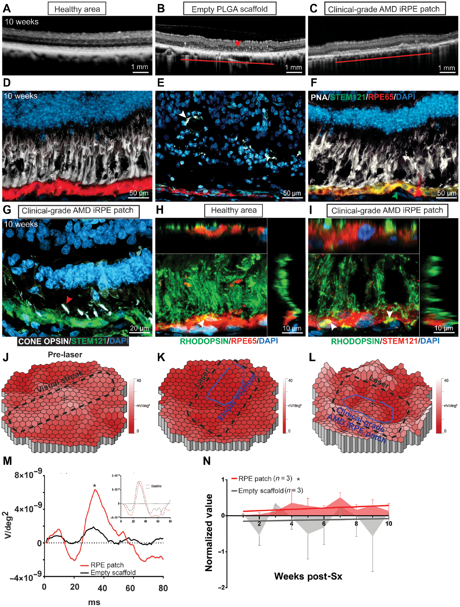
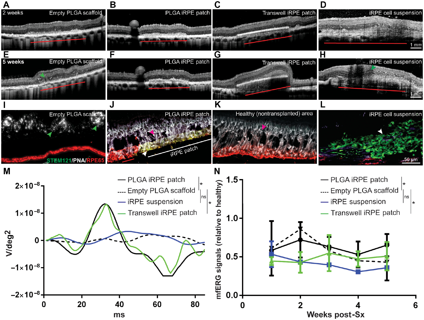
Considerable progress has been made in testing stem cell-derived retinal pigment epithelium (RPE) as a potential therapy for age-related macular degeneration (AMD). However, the recent reports of oncogenic mutations in induced pluripotent stem cells (iPSCs) underlie the need for robust manufacturing and functional validation of clinical-grade iPSC-derived RPE before transplantation. Here, we developed oncogenic mutation-free clinical-grade iPSCs from three AMD patients and differentiated them into clinical-grade iPSC-RPE patches on biodegradable scaffolds. Functional validation of clinical-grade iPSC-RPE patches revealed specific features that distinguished transplantable from nontransplantable patches. Compared to RPE cells in suspension, our biodegradable scaffold approach improved integration and functionality of RPE patches in rats and in a porcine laser-induced RPE injury model that mimics AMD-like eye conditions. Our results suggest that the in vitro and in vivo preclinical functional validation of iPSC-RPE patches developed here might ultimately be useful for evaluation and optimization of autologous iPSC-based therapies.


Conflict of interest statement

Competing interests: K.B., A.M., S.M. are inventors on patent application - Method for generating retinal pigment epithelium (rpe) cells from induced pluripotent stem cells (ipscs) (W0201412077A2); K.B., L.C., B.J.S are inventors on patent application - Method for reproducible differentiation of clinical-grade retinal pigment epithelium cells (W02017044483A1); L.C. and C.S. are inventors on patent application - Macs-based purification of stem cell-derived retinal pigment epithelium (US20170067017A1). K.B., V.K., S.C., J.A., and A.M. are inventors on patent application - Tissue clamp and implantation method (W02018089521A1); A.M. is an inventor on patent application - Surgical tool and method for ocular tissue transplantation (US20170128263A1); All other authors declare that they have no competing interests.

Figures

Fig. 1.
Fig. 1.. Generation of clinical-grade iPSC-RPE cells
(A) Workflow illustrating a pipeline to manufacture and test autologous clinical-grade iPSC-RPE-patches with the goal of filing a phase I clinical trial Investigational New Drug (IND)-application to the FDA. (B) Time-line of clinical-grade iRPE differentiation. Clinical-grade iRPE differentiation takes 77 days, is initiated with monolayer iPSCs and performed using xeno-free reagents. Neuro Ectoderm Induction Medium (NEIM); RPE Induction Medium (RPEIM); RPE Commitment Medium (RPECM); RPE Growth Medium (RPEGM); RPE Maturation Medium (RPEMM). (C) Coding-region sequencing of 223 oncogenes at 2000x depth for all nine clinical-grade AMD iPSC clones. (D, E) Flow cytometry analysis of clinical-grade iPSC-RPE derived from three AMD patients, performed at the RPE progenitor stage day (D)17, RPE-commitment stage (D27), and immature RPE-stage (D42) (n=6). Analysis of variance (ANOVA) was performed to determine changes in percent positive cells; ***p=0.0001 for PAX6/MITF and ***p=8.9×10⁻¹⁴ for MITF; Dunn’s test was performed for pair-wise comparisons; p-values: 2B/2C- 3C/3D=0.909; 2B/2C-4B/4C=0.400; 3C/3D-4B/4C=0.319 (F) RPE-specific gene expression from D5–D42 of clinical-grade iPSC-RPE differentiation (n=6). Dunn’s test was performed for pair-wise comparisons; p-values: 2B/2C- 3C/3D=0.721; 2B/2C- 4B/4C=0.719; 3C/3D-4B/4C=0.999.
Fig. 2.
Fig. 2.. Generation of functionally mature AMD iRPE-patche
(A) Young’s Modulus of different PLGA-scaffolds. Two-tailed t-test; **p<0.01 (B) SEM showing surface topology of single-layer fused PLGA scaffold. (C) Representative immunostaining for mature-RPE marker RPE65 (red) and human-specific antigen STEM121 (green), top panel; RPE-pigmentation protein GPNMB (red) and Bruch’s membrane protein COLLAGEN IV (green), middle panel; and COLLAGEN VIII, Bruch’s membrane marker (red), bottom panel (n=3). (D) Representative TEM of iRPE-patch on transwell membrane or the PLGA-scaffold. Basal infoldings can be seen in the case of PLGA scaffold (inset) (n=3). (E) ΔcT values of RPE-specific genes are displayed for all eight iRPE-patches from three AMD donors (n=8). Dunn’s test was performed to determine pair-wise comparisons; p-values: 2B/2C- 3C/3D=0.999; 2B/2C-4B/4C=0.150; 3C/3D-4B/4C=0.094. (F) Live TER measurement during the last three weeks (D54–77) of iRPE-patch maturation. Representative data from three clones (3A, 3D, 4A) are displayed (n=8). Dunn’s test was performed to determine changes in TER overtime; p-values: 3A-3D=0.630; 3A-4A=0.845; 3D-4A=0.968 (G) Graphs shows phagocytosis ratio for t AMD-iRPE-patches (n=8). Dunn’s test was performed to compare iRPE from different donors; p-values: 2B/2C-3A/3C/3D=0.005; 2B/2C-4A/4B/4C=0.395; 3A/3C/3D −4A/4B/4C = 0.63. (H, I) Principle Component Analysis (PCA) combining data from the following assays (morphometric, gene expression, TER, and phagocytosis) showing variation between clones across PC1. PCA was performed based on k-nearest neighbors and bootstrap hierarcial clustering was performed to determine differences between iRPE samples; *p<0.05. (H). PCA plotted without D2B (I) (n=7–8).
Fig. 3.
Fig. 3.. Safey and efficacy assessment of clinical-grade AMD iRPE-patch in rodent models
(A-C) Representative En face infrared image (A), OCT (B), and immunohistochemistry for human antigen (STEM121 – red, C) showing sub-retinal location and integration of the 0.5 mm diameter clinical-grade AMD-iRPE-patch (red arrowhead). Black arrowhead marks rat RPE cells – see inset for higher magnification) in the sub-retinal space of immunocompromised rat at ten weeks post-surgery. (n=20) (D) Represenative immunohistochemistry for STEM121 – purple (red arrowhead, see inset for higher magnifications) confirms the presence of clinical-grade AMD-iRPE cells injected in rat eye. Note, purple color in photoreceptor outer segments is due to hematoxylin stain. Rat RPE are not positive for STEM121 (black arrowhead; see inset for higher magnification). (E) Representative STEM121 -red immunostaining (red arrowhead) showing integration of a small number of human cells in the rat RPE (black arrowhead; see inset for higher magnification) (n=10). (F) Representative Ki67 immunostaining showing lack of positivity. Human cells are indicated by red arrowhead (see inset for higher magnification; rat RPE is marked with black arrowhead). (G, I) Representative photomontage of RCS rat retina showing outer nuclear layer (ONL, arrowheads) with transplanted iPSC-RPE-patch (~10,000 cells on a 1 mm diameter patch) (G) or iRPE cell suspension (100,000 cells) (I), compared with the degenerated ONL in non-transplanted areas (arrow, n=10). (H, J) Representative immunofluorescence staining of iRPE-patch (H) or iRPE cell suspension (J) implanted retina with ONL rescue (arrowheads) (red – HuNu, human nuclear antigen, green; human-specific anti-PMEL17). Note, red arrowhead in (H) points to iRPE cells that likely dislodged from the scaffold during transplantation. (K) Optokinetic tracking thresholds at P90. (n = 10).; *p<0.05, **p<0.001 determined using ANOVA analysis.
Fig. 4.
Fig. 4.. Development of a porcine iRPE-patch efficacy model
(A) Schematics of micropulse laser injuring the pig RPE; insert, fluorescein angiogram depicting laser-induced outer blood-retinal-barrier breakdown. (B, C) Representative OCT images at 24 h and 48 h post laser (arrowheads indicates RPE-thinning), n=3. (D) Heatmap of the P1 values of the visual streak region after 1% or 3% duty cycle laser (laser areas outlined with dashed lines in). White-red indicates the highest P1 values and blue indicate the lowest. (E) Average mfERG waveform from healthy (black), 1% (light green) and 3% (dark green) duty cycle laser areas. (F-I) Representative immunostaining for TUNEL (Green), RPE65 (Yellow), and PNA (magenta) (F, G) and H&E staining (H, I) at 48 hours (arrowheads indicate apoptotic RPE), n=3.
Fig. 5.
Fig. 5.. Efficacy assessment of clinical-grade AMD iRPE-patch in a porcine retinal degeneration model.
(A-C) Comparison of OCT from retina over a healthy region, retina transplanted with an empty PLGA scaffold, or a retina transplanted with clinical-grade AMD iRPE-patch (horizontal lines) n = 3. (D-F) Immunostaining for STEM 121 (green, arrowhead, F) and RPE65 (red) in the pig eye. PNA staining is shown in white; white arrowhead in E marks retinal tubulations) n = 3. (G) Immunostaining for Red, Blue, and Green cone opsins (white; red arrowhead) and STEM121(green) in the pig eye after iRPE-patch transplantation. (H, I) Rhodopsin (green) immunostaining shows phagocytosed (white arrowheads) photoreceptor outer segments (POS) by healthy pig RPE immunostained with RPE65 (red) and by human iRPE cells immunostained with STEM121 (red). Z-sections show POS localization inside pig and human RPE cells n = 3. (J-L) Heat maps of N1P1 mfERG responses. (M, N) Average mfERG waveform (M), and mfERG data over 10 weeks of follow up post-surgery (N) n=3. LME was performed for data analysis and ANOVA to determine statistical significance of the data *p<0.05.
Fig. 6.
Fig. 6.. Integration of iRPE-patch in a laser-induced retinal degeneration porcine model.
(A-H) Representative OCT images of pig eyes two weeks and five weeks after transplantion with empty scaffold, PLGA iRPE-patch, transwell iRPE-patch, and iRPE cell suspension. iRPE-transplants are indicated by red horizontal line. Green arrowheads point to retinal tubulations. (I-L) Representative immunostaining for human antigen STEM121 (green) and RPE65 (red) in the pig eye (iRPE-patch is indicated by horizontal white line in J; white arrowhead in J and L marks iRPE-transplants; green arrowhead in I marks retinal tubulations; and red arrowhead in J and K marks rat photoreceptors). n=3. (M, N) Individual and average mfERG responses from different transplant conditions (n=3; *p<0.05 determined using ANOVA).

References

    1. Fischbach MA, Bluestone JA, Lim WA, Cell-based therapeutics: the next pillar of medicine. Sci Transl Med 5, 177–179 (2013). - PMC - PubMed
    1. Song MJ, Bharti K, Looking into the future: Using induced pluripotent stem cells to build two and three dimensional ocular tissue for cell therapy and disease modeling. Brain Res 1638, 2–14 (2016). - PMC - PubMed
    1. Joussen AM et al., Autologous translocation of the choroid and retinal pigment epithelium in patients with geographic atrophy. Ophthalmology 114, 551–560 (2007). - PubMed
    1. van Zeeburg EJ, Maaijwee KJ, Missotten TO, Heimann H, van Meurs JC, A free retinal pigment epithelium-choroid graft in patients with exudative age-related macular degeneration: results up to 7 years. Am J Ophthalmol 153, 120–127.e122 (2012). - PubMed
    1. Schwartz SD et al., Embryonic stem cell trials for macular degeneration: a preliminary report. Lancet 379, 713–720 (2012). - PubMed

Publication types