. 2019 Mar 13;39(11):2080-2090.
doi: 10.1523/JNEUROSCI.2701-18.2019. Epub 2019 Jan 16.

Prefrontal Corticotropin-Releasing Factor (CRF) Neurons Act Locally to Modulate Frontostriatal Cognition and Circuit Function

Affiliations

Prefrontal Corticotropin-Releasing Factor (CRF) Neurons Act Locally to Modulate Frontostriatal Cognition and Circuit Function

Sofiya Hupalo et al. J Neurosci. .

Abstract

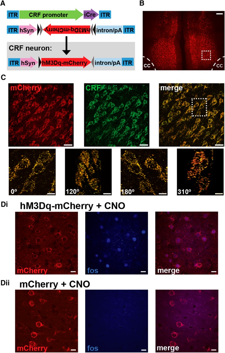
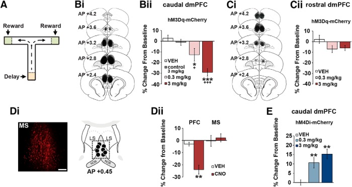
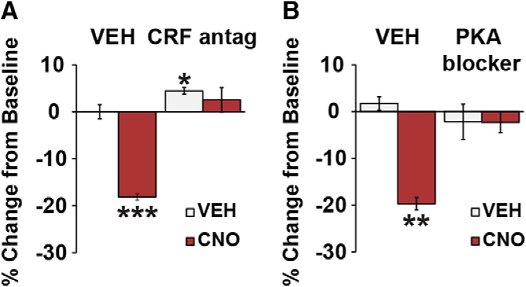
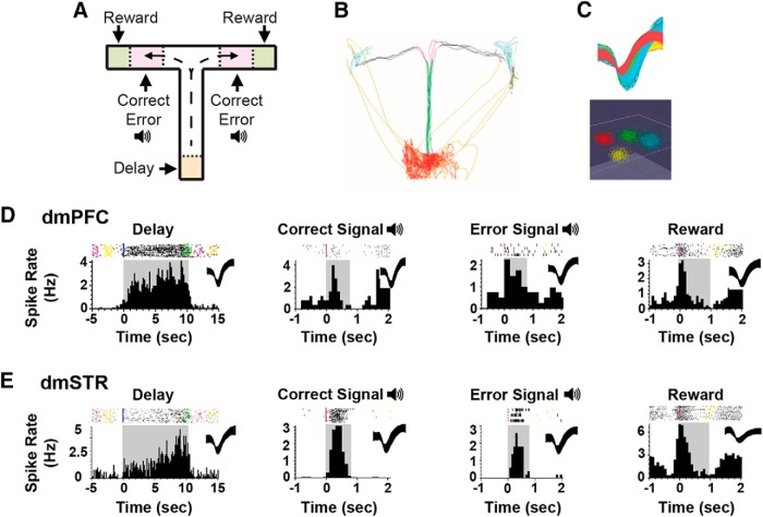
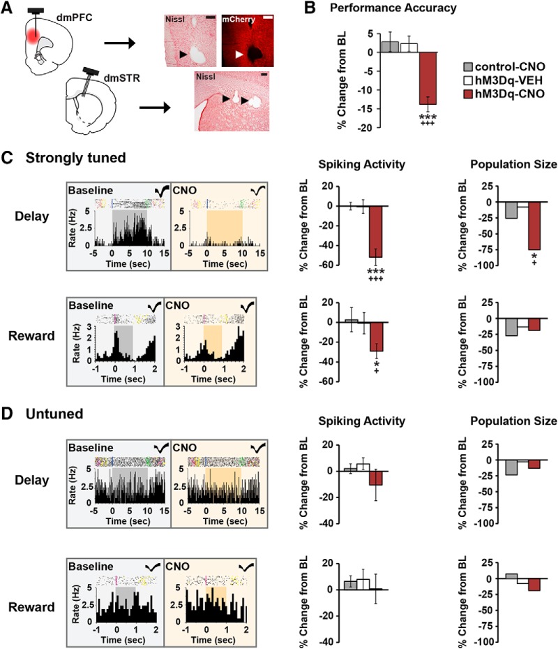
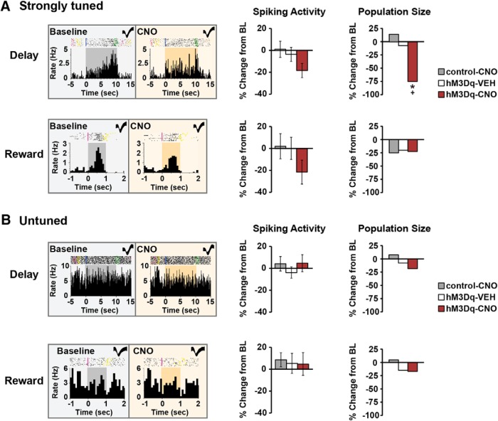
The PFC and extended frontostriatal circuitry support higher cognitive processes that guide goal-directed behavior. PFC-dependent cognitive dysfunction is a core feature of multiple psychiatric disorders. Unfortunately, a major limiting factor in the development of treatments for PFC cognitive dysfunction is our limited understanding of the neural mechanisms underlying PFC-dependent cognition. We recently demonstrated that activation of corticotropin-releasing factor (CRF) receptors in the caudal dorsomedial PFC (dmPFC) impairs higher cognitive function, as measured in a working memory task. Currently, there remains much unknown about CRF-dependent regulation of cognition, including the source of CRF for cognition-modulating receptors and the output pathways modulated by these receptors. To address these issues, the current studies used a viral vector-based approach to chemogenetically activate or inhibit PFC CRF neurons in working memory-tested male rats. Chemogenetic activation of caudal, but not rostral, dmPFC CRF neurons potently impaired working memory, whereas inhibition of these neurons improved working memory. Importantly, the cognition-impairing actions of PFC CRF neurons were dependent on local CRF receptors coupled to protein kinase A. Additional electrophysiological recordings demonstrated that chemogenetic activation of caudal dmPFC CRF neurons elicits a robust degradation of task-related coding properties of dmPFC pyramidal neurons and, to a lesser extent, medium spiny neurons in the dorsomedial striatum. Collectively, these results demonstrate that local CRF release within the caudal dmPFC impairs frontostriatal cognitive and circuit function and suggest that CRF may represent a potential target for treating frontostriatal cognitive dysfunction.SIGNIFICANCE STATEMENT The dorsomedial PFC and its striatal targets play a critical role in higher cognitive function. PFC-dependent cognitive dysfunction is associated with many psychiatric disorders. Although it has long-been known that corticotropin-releasing factor (CRF) neurons are prominent within the PFC, their role in cognition has remained unclear. Using a novel chemogenetic viral vector system, the present studies demonstrate that PFC CRF neurons impair working memory via activation of local PKA-coupled CRF receptors, an action associated with robust degradation in task-related frontostriatal neuronal coding. Conversely, suppression of constitutive PFC CRF activity improved working memory. Collectively, these studies provide novel insight into the neurobiology of cognition and suggest that CRF may represent a novel target for the treatment of cognitive dysfunction.

Keywords: DREADDs; corticotropin-releasing factor; in vivo electrophysiology; prefrontal cortex; striatum; working memory.


Figures

Figure 1.
Figure 1.
Chemogenetic approach. A, Schematic depicting dual viral system to activate (CRF-Cre + hSyn-DIO-hM3Dq) or inhibit (CRF-Cre + hSyn-DIO-hM4Di) CRF neurons in the PFC. B, Photomicrograph depicting mCherry expression in the caudal dmPFC from a CRF-hM3Dq-treated animal. Scale bar, 200 μm. cc, Corpus callosum. C, Top, Collapsed 30 μm z stack from inset in B, demonstrating mCherry colocalization with CRF-ir cells. Scale bar, 30 μm. Bottom, Inset from above, rotated at various angles. Scale bar, 10 μm. Di, CNO elicits an excitatory influence in PFC CRF neurons as measured by Fos-ir in mCherry neurons of CNO-treated CRF-hM3Dq animals. Dii, Absence of Fos-ir in the PFC of CNO-treated viral controls (mCherry + CNO; CRF-Cre hSyn-DIO-mCherry). Scale bar, 10 μm.
Figure 2.
Figure 2.
CRF neurons in the caudal, but not rostral, dmPFC modulate working memory. A, T-maze schematic. Bi, Schematics depict hM3Dq viral spread in the caudal dmPFC (anteroposterior 3.2–2.2) from all animals tested. Bii, CNO dose-dependently impairs task performance relative to vehicle (n = 7) and CNO-treated viral control animals (n = 7; control 3 mg/kg). Ci, hM3Dq viral spread in the rostral dmPFC (anteroposterior 4.2–3.2). Cii, Chemogenetic activation of CRF neurons in the rostral dmPFC has no significant effects on task performance relative to vehicle (n = 6) and viral controls (n = 7). Di, Left, Retrograde mCherry cell body labeling observed in the MS in ∼30% of animals. Right, Schematics representing intra-MS infusion sites (n = 4). Dii, When infused into the PFC, 0.5 mm CNO robustly impairs task performance (n = 7), while having no effects on performance when infused into the MS (n = 4). E, Chemogenetic suppression of CRF neurons in the caudal dmPFC improves task performance relative to vehicle (n = 8) and CNO-treated viral controls (n = 7). Results are mean ± SEM percentage change in accuracy relative to baseline. *p < 0.05 versus vehicle. +p < 0.001 versus viral controls. **p < 0.01 versus vehicle. ***p < 0.001 versus vehicle. +++p < 0.001 versus viral controls.
Figure 3.
Figure 3.
Working memory actions of caudal dmPFC CRF neurons are dependent on local CRF receptors coupled to PKA. A, Intra-PFC infusion of the nonselective CRF antagonist d-Phe-CRF (100 ng/hemisphere) blocks the working memory-impairing effects of chemogenetic activation of caudal dmPFC CRF neurons (3 mg/kg CNO; n = 9). B, Intra-PFC infusion of the PKA inhibitor Rp-cAMPs (20 nm/hemisphere) also blocks the working memory-impairing effects of 3 mg/kg CNO (n = 5). Results are mean ± SEM percentage change in accuracy relative to baseline. *p < 0.05. **p < 0.01. ***p < 0.001.
Figure 4.
Figure 4.
Frontostriatal recordings in working memory-tested rats. A, T-maze schematic illustrating task events, including delay, two distinct auditory tones serving as outcome-related signals on correct versus error trials, and sugar reward. B, Animal position is tracked using video recordings and infrared beams. C, Top, Action potential waveforms of 4 discriminated WS dmPFC neurons. Bottom, Waveforms from these units exhibit separable clusters in 3D-principal component space. D, Exemplar rasters (top) and PETHs (bottom) of WS dmPFC neurons exhibiting punctate excitatory responses during T-maze events. Shaded areas of PETHs represent duration of interval. Spiking rates were calculated for 100 ms time bins. E, Exemplar rasters and PETHs of putative MSNs within the dmSTR displaying strong task tuning.
Figure 5.
Figure 5.
Chemogenetic activation of CRF neurons in the caudal dmPFC degrades delay and reward signaling in WS dmPFC neurons. A, Left, Schematics depicting CRF-hM3Dq expression in the caudal dmPFC and electrode placements in the dmPFC and dmSTR. Right, 4× photomicrographs depicting placement of one electrode in layer V of the dmPFC and three electrodes in the dmSTR. Scale bar, 250 μm. B, In recorded animals, CNO impaired task performance in the hM3Dq group (5 animals, 7 recording sessions), but not in vehicle (4 animals, 10 sessions) or CNO-treated viral controls (3 animals, 10 recording sessions). C, Left, Exemplar rasters/PETHs demonstrating task-related activity of strongly tuned delay (top) and reward (bottom) WS neurons under baseline and CNO conditions (delay, 10 s; reward, 1 s). Middle, CNO-induced activation of dmPFC CRF neurons robustly suppressed task-related activity of strongly tuned delay (n = 24) and reward (n = 16), WS neurons relative to vehicle (delay, n = 25; reward, n = 15), and CNO-treated viral controls (delay, n = 54; reward, n = 11). Right, PFC CRF neuronal activation diminished the population size of strongly tuned delay (top), but not reward (bottom) neurons. D, Left, Exemplar rasters/PETHs of a WS neuron untuned to delay (top) and reward (bottom) under baseline and CNO conditions. CNO had no significant effects on task-related activity (middle) or population sizes (right) of these neurons in hM3Dq animals (delay, n = 55; reward, n = 65), relative to vehicle (delay, n = 40; reward, n = 59), and viral controls (delay, n = 64; reward, n = 92). *p < 0.05 versus control-CNO. +p < 0.05 versus hM3Dq-SAL. ***p < 0.001 versus control-CNO. +++p < 0.001 versus hM3Dq-SAL.
Figure 6.
Figure 6.
Effects of PFC CRF neuronal activation on task-related activity of dmSTR MSNs. A, Left, Exemplar rasters/PETHs demonstrating task-related activity of strongly tuned delay (top) and reward (bottom) MSNs under baseline and CNO conditions. Middle, CNO elicited a trend for suppression of task-related activity of strongly tuned delay (n = 12) and reward MSNs (n = 9) in hM3Dq animals that was not observed with vehicle (delay, n = 28; reward, n = 15) or CNO-treated viral controls (delay, n = 28; reward, n = 12). Right, PFC CRF neuronal activation diminished the population size of strongly tuned delay (top), but not reward (bottom), MSNs. B, Left, Exemplar rasters/PETHs of MSNs untuned to delay (top) and reward (bottom) MSNs. CNO elicited no significant effects on task-related activity (middle) or the population size (right) of untuned MSNs in hM3Dq animals (delay, n = 27; reward, n = 35), vehicle-treated hM3Dq animals (delay, n = 26; reward, n = 65), or CNO-treated viral controls (delay, n = 39; reward, n = 61). *p < 0.05 versus control-CNO. +p < 0.05 versus hM3Dq-SAL.

References

    1. Akhlaghpour H, Wiskerke J, Choi JY, Taliaferro JP, Au J, Witten IB (2016) Dissociated sequential activity and stimulus encoding in the dorsomedial striatum during spatial working memory. eLife 5:e19507. 10.7554/eLife.19507 - DOI - PMC - PubMed
    1. Alsene KM, Rajbhandari AK, Ramaker MJ, Bakshi VP (2011) Discrete forebrain neuronal networks supporting noradrenergic regulation of sensorimotor gating. Neuropsychopharmacology 36:1003–1014. 10.1038/npp.2010.238 - DOI - PMC - PubMed
    1. Arnsten AF. (2009) Stress signalling pathways that impair prefrontal cortex structure and function. Nat Rev Neurosci 10:410–422. 10.1038/nrn2648 - DOI - PMC - PubMed
    1. Arnsten AF. (2011) Catecholamine influences on dorsolateral prefrontal cortical networks. Biol Psychiatry 69:e89–e99. 10.1016/j.biopsych.2011.01.027 - DOI - PMC - PubMed
    1. Batuev AS, Kursina NP, Shutov AP (1990) Unit activity of the medial wall of the frontal cortex during delayed performance in rats. Behav Brain Res 41:95–102. 10.1016/0166-4328(90)90145-5 - DOI - PubMed

Publication types

Substances

LinkOut - more resources