Representation of Auditory Motion Directions and Sound Source Locations in the Human Planum Temporale
- PMID: 30651333
- PMCID: PMC6433766
- DOI: 10.1523/JNEUROSCI.2289-18.2018
Representation of Auditory Motion Directions and Sound Source Locations in the Human Planum Temporale
Erratum in
-
Erratum: Battal et al., "Representation of Auditory Motion Directions and Sound Source Locations in the Human Planum Temporale".J Neurosci. 2019 Jul 10;39(28):5627. doi: 10.1523/JNEUROSCI.0996-19.2019. Epub 2019 May 13. J Neurosci. 2019. PMID: 31085609 Free PMC article. No abstract available.
Abstract
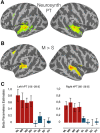
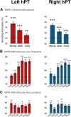
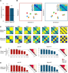
The ability to compute the location and direction of sounds is a crucial perceptual skill to efficiently interact with dynamic environments. How the human brain implements spatial hearing is, however, poorly understood. In our study, we used fMRI to characterize the brain activity of male and female humans listening to sounds moving left, right, up, and down as well as static sounds. Whole-brain univariate results contrasting moving and static sounds varying in their location revealed a robust functional preference for auditory motion in bilateral human planum temporale (hPT). Using independently localized hPT, we show that this region contains information about auditory motion directions and, to a lesser extent, sound source locations. Moreover, hPT showed an axis of motion organization reminiscent of the functional organization of the middle-temporal cortex (hMT+/V5) for vision. Importantly, whereas motion direction and location rely on partially shared pattern geometries in hPT, as demonstrated by successful cross-condition decoding, the responses elicited by static and moving sounds were, however, significantly distinct. Altogether, our results demonstrate that the hPT codes for auditory motion and location but that the underlying neural computation linked to motion processing is more reliable and partially distinct from the one supporting sound source location.SIGNIFICANCE STATEMENT Compared with what we know about visual motion, little is known about how the brain implements spatial hearing. Our study reveals that motion directions and sound source locations can be reliably decoded in the human planum temporale (hPT) and that they rely on partially shared pattern geometries. Our study, therefore, sheds important new light on how computing the location or direction of sounds is implemented in the human auditory cortex by showing that those two computations rely on partially shared neural codes. Furthermore, our results show that the neural representation of moving sounds in hPT follows a "preferred axis of motion" organization, reminiscent of the coding mechanisms typically observed in the occipital middle-temporal cortex (hMT+/V5) region for computing visual motion.
Keywords: auditory motion; direction selectivity; fMRI; multivariate analyses; planum temporale; spatial hearing.
Copyright © 2019 the authors 0270-6474/19/392208-13$15.00/0.
Figures


Similar articles
-
Direct Structural Connections between Auditory and Visual Motion-Selective Regions in Humans.J Neurosci. 2021 Mar 17;41(11):2393-2405. doi: 10.1523/JNEUROSCI.1552-20.2021. Epub 2021 Jan 29. J Neurosci. 2021. PMID: 33514674 Free PMC article.
-
Structural and Functional Network-Level Reorganization in the Coding of Auditory Motion Directions and Sound Source Locations in the Absence of Vision.J Neurosci. 2022 Jun 8;42(23):4652-4668. doi: 10.1523/JNEUROSCI.1554-21.2022. Epub 2022 May 2. J Neurosci. 2022. PMID: 35501150 Free PMC article.
-
Active Sound Localization Sharpens Spatial Tuning in Human Primary Auditory Cortex.J Neurosci. 2018 Oct 3;38(40):8574-8587. doi: 10.1523/JNEUROSCI.0587-18.2018. Epub 2018 Aug 20. J Neurosci. 2018. PMID: 30126968 Free PMC article.
-
Psychophysics and neuronal bases of sound localization in humans.Hear Res. 2014 Jan;307:86-97. doi: 10.1016/j.heares.2013.07.008. Epub 2013 Jul 22. Hear Res. 2014. PMID: 23886698 Free PMC article. Review.
-
Distributed coding of sound locations in the auditory cortex.Biol Cybern. 2003 Nov;89(5):341-9. doi: 10.1007/s00422-003-0439-1. Epub 2003 Nov 12. Biol Cybern. 2003. PMID: 14669014 Review.
Cited by
-
Multimodal processing in face-to-face interactions: A bridging link between psycholinguistics and sensory neuroscience.Front Hum Neurosci. 2023 Feb 2;17:1108354. doi: 10.3389/fnhum.2023.1108354. eCollection 2023. Front Hum Neurosci. 2023. PMID: 36816496 Free PMC article.
-
Electrocorticography Evidence of Tactile Responses in Visual Cortices.Brain Topogr. 2020 Sep;33(5):559-570. doi: 10.1007/s10548-020-00783-4. Epub 2020 Jul 13. Brain Topogr. 2020. PMID: 32661933 Free PMC article.
-
Direct Structural Connections between Auditory and Visual Motion-Selective Regions in Humans.J Neurosci. 2021 Mar 17;41(11):2393-2405. doi: 10.1523/JNEUROSCI.1552-20.2021. Epub 2021 Jan 29. J Neurosci. 2021. PMID: 33514674 Free PMC article.
-
Do you hear what I see? How do early blind individuals experience object motion?Philos Trans R Soc Lond B Biol Sci. 2023 Jan 30;378(1869):20210460. doi: 10.1098/rstb.2021.0460. Epub 2022 Dec 13. Philos Trans R Soc Lond B Biol Sci. 2023. PMID: 36511418 Free PMC article. Review.
-
Intelligibility of audiovisual sentences drives multivoxel response patterns in human superior temporal cortex.Neuroimage. 2022 Feb 15;247:118796. doi: 10.1016/j.neuroimage.2021.118796. Epub 2021 Dec 11. Neuroimage. 2022. PMID: 34906712 Free PMC article.
References
-
- Ahveninen J, Jaaskelainen IP, Raij T, Bonmassar G, Devore S, Hämäläinen M, Levänen S, Lin FH, Sams M, Shinn-Cunningham BG, Witzel T, Belliveau JW (2006) Task-modulated “what” and “where” pathways in human auditory cortex. Proc Natl Acad Sci U S A 103:14608–14613. 10.1073/pnas.0510480103 - DOI - PMC - PubMed
Publication types
MeSH terms
LinkOut - more resources
Full Text Sources
