. 2019 Mar 8;294(10):3794-3805.
doi: 10.1074/jbc.RA118.005419. Epub 2019 Jan 16.

Biochemical and structural analyses suggest that plasminogen activators coevolved with their cognate protein substrates and inhibitors

Affiliations

Biochemical and structural analyses suggest that plasminogen activators coevolved with their cognate protein substrates and inhibitors

Agnieszka Jendroszek et al. J Biol Chem. .

Abstract

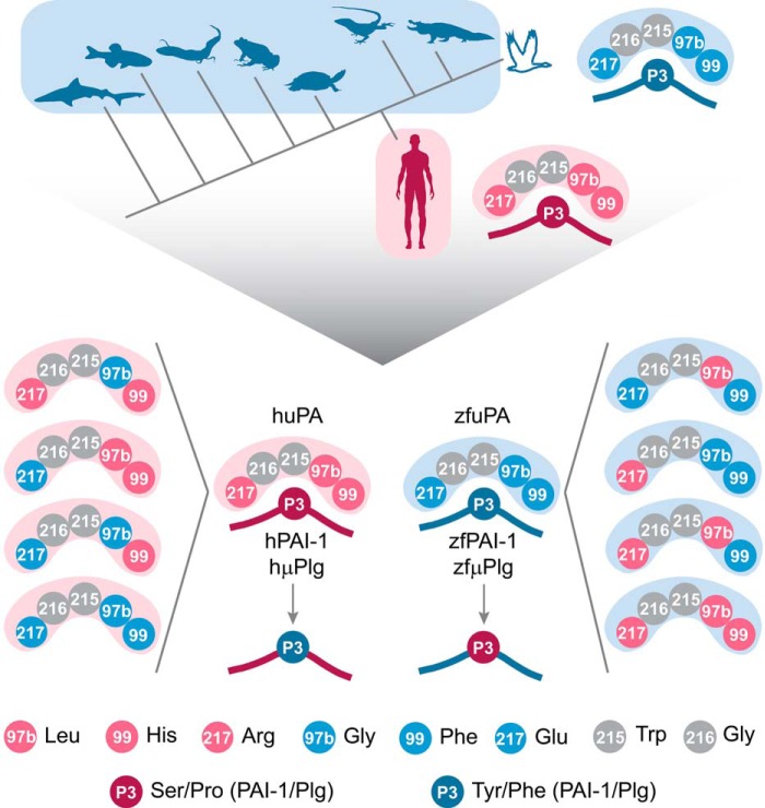
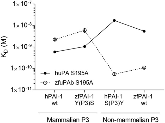
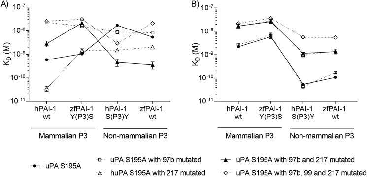
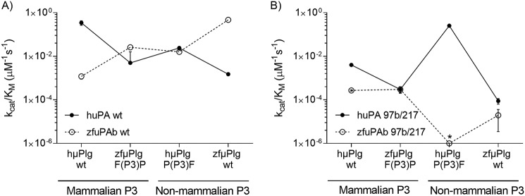
Protein sequences of members of the plasminogen activation system are present throughout the entire vertebrate phylum. This important and well-described proteolytic cascade is governed by numerous protease-substrate and protease-inhibitor interactions whose conservation is crucial to maintaining unchanged protein function throughout evolution. The pressure to preserve protein-protein interactions may lead to either co-conservation or covariation of binding interfaces. Here, we combined covariation analysis and structure-based prediction to analyze the binding interfaces of urokinase (uPA):plasminogen activator inhibitor-1 (PAI-1) and uPA:plasminogen complexes. We detected correlated variation between the S3-pocket-lining residues of uPA and the P3 residue of both PAI-1 and plasminogen. These residues are known to form numerous polar interactions in the human uPA:PAI-1 Michaelis complex. To test the effect of mutations that correlate with each other and have occurred during mammalian diversification on protein-protein interactions, we produced uPA, PAI-1, and plasminogen from human and zebrafish to represent mammalian and nonmammalian orthologs. Using single amino acid point substitutions in these proteins, we found that the binding interfaces of uPA:plasminogen and uPA:PAI-1 may have coevolved to maintain tight interactions. Moreover, we conclude that although the interaction areas between protease-substrate and protease-inhibitor are shared, the two interactions are mechanistically different. Compared with a protease cleaving its natural substrate, the interaction between a protease and its inhibitor is more complex and involves a more fine-tuned mechanism. Understanding the effects of evolution on specific protein interactions may help further pharmacological interventions of the plasminogen activation system and other proteolytic systems.

Keywords: protein evolution; serine protease; serpin; substrate specificity; surface plasmon resonance (SPR).


Conflict of interest statement

The authors declare that they have no conflicts of interest with the contents of this article

Figures

Figure 1.
Figure 1.
Michaelis complex between uPA and PAI-1. A, structure of Michaelis complex between human PAI-1 (yellow) and uPA (white) (PDB ID 3PB1). B, in complexes with uPA, main chains of the P1 to P3 residues of PAI-1 (yellow) align with the P1–P3 residues of EGR-cmk (green) (PDB ID 1LMW). C, P3 Ser of hPAI-1 forms polar interactions with the S3-pocket forming residues of huPA (Leu-97b, His-99, Gly-216). Hydrogen bonds are shown as dashed lines. D and E, charge and size of the S3-pocket in huPA and zfuPA are different. Enlarged S3-pocket of zfuPA can accommodate bulky P3 residues of zfPAI-1 (cyan). Structure of the zfuPA:zfPAI-1 Michaelis complex was modeled in SwissModel using a human uPA:PAI-1 Michaelis complex structure as the template (PDB ID 3PB1). The RCL of zfPAI-1 is shown in cyan, with P1 to P3 residues shown as sticks.
Figure 2.
Figure 2.
Multiple sequence alignment of the PAI-1 RCL (A), plasminogen activation loop (C), and residues forming 97b- and 217-loops in uPA (B) and tPA (D). To compare uPA, tPA, plasminogen, and PAI-1 from various vertebrates, sequences of each monophyletic group were aligned in ClustalOmega and a consensus was determined. Numbers in the brackets refer to the number of sequences used for consensus determination. Ambiguous resides are shown as X.
Figure 3.
Figure 3.
Strategy for mutagenesis of uPA, PAI-1, and μPlg from zebrafish and human. Based on multiple sequence alignment and MI calculations, correlated mutations between the S3-pocket–lining residues of uPA and the P3 residue of PAI-1 and S3-pocket residues of uPA and P3 residue of plasminogen were detected. The uPA S3 specificity pocket is composed of residues 97b, 99, 215, 216, and 217 of which residues Trp-215 and Gly-216 (marked in gray) are conserved through the vertebrate phylum. Residues 97b, 99, and 217 are Leu, His, and Arg in mammalian uPA (pink) and Gly, Phe, and Glu in uPA from nonmammalian species (blue). The P3 residue of PAI-1 inserts into the S3 specificity pocket of uPA and forms polar contacts with the S3-pocket–lining residues (16). The P3 residue is either Ser in mammalian PAI-1 (dark pink) or Tyr in nonmammalian PAI-1 (dark blue). The P3 residue of plasminogen is either Pro (dark pink) or Phe (dark blue) in mammalian and nonmammalian proteins, respectively. As correlated mutations occurred after diversification of mammals, we produced human (pink) and zebrafish (blue) uPA, PAI-1, and plasminogen, representing mammalian and nonmammalian orthologues, respectively. We then exchanged the residues of interest alone or in combination and tested if interactions between uPA:PAI-1 and uPA:plasminogen were affected.
Figure 4.
Figure 4.
SPR analysis of uPA:PAI-1 Michaelis complex formation. To test the importance of the P3 residue of PAI-1 on species specificity in reaction with uPA, the P3 residues were exchanged between hPAI-1 and zfPAI-1. hPAI-1 WT, zfPAI-1 WT, hPAI-1 S(P3)Y, and zfPAI-1 Y(P3)S variants were tested for binding to huPA S195A (closed symbols) and zfuPA S195A (open symbols) captured on the surface of the SPR chip via specific antibody. The S195A mutation in the protease enabled the inhibitory reaction to stop at the Michaelis complex formation step. Each point represents the mean value for three individual KD determinations with standard deviation. Helper lines interconnecting the individual data point are given as visual trend guides. Representative sensorgrams with the fit are shown in Fig. S2.
Figure 5.
Figure 5.
SPR-based screening of uPA mutants. Importance of the S3-pocket–lining residues of uPA on species specificity was tested by exchanging the residues alone or simultaneously between huPA S195A and zfuPA S195A. Created huPA variants: (A) huPA L97bG/S195A, huPA S195A/R217E, huPA L97bG/S195A/R217E, huPA L97bG/H99F/S195A/R217E, and zfuPA variants: (B) zfuPA G97bL/S195A, zfuPA S195A/E217R, zfuPA G97bL/S195A/E217R, and zfuPA G97bL/F99H/S195A/E217R were captured on the surface of the SPR chip via a specific antibody and tested for binding to hPAI-1 WT, hPAI-1 S(P3)Y, zfPAI-1 WT and zfPAI-1 Y(P3)S. Each point represents the mean of three independent KD determinations with standard deviation. Helper lines interconnecting the individual data point are given as visual trend guides. Representative sensorgrams with the fit are shown in Fig. S2.
Figure 6.
Figure 6.
Activation of μPlg by WT (A) and 97b/217 mutants (B) of huPA and zfuPA measured by coupled assay. A, importance of the P3 residue of plasminogen on species specificity was tested by exchanging the P3 residues between hμPlg and zfμPlg followed by determination of activation rates of hμPlg WT, hμPlg P(P3)F, zfμPlg WT, and zfμPlg F(P3)P by huPA WT (closed symbols) and zfuPA WT (open symbols). B, because the huPA L97bG/S195A/R217E showed reversed specificity toward the P3 residue of PAI-1 compared with huPA S195A, we have chosen huPA L97bG/R217E (closed symbols) and zfuPA G97bL/E217R (open symbols) and tested for activation of hμPlg and zfμPlg variants. The activation rate of zfμPlg F(P3)P by zfuPA G97bL/E217R (marked with *) was too slow to be measured by this method. To eliminate the effect of the plasminogen conformational state on activation rate and to focus on the active site interactions, μPlg was used instead of full-length plasminogen. Each point represents the mean value calculated from three individual kcat/Km determinations with standard deviation. Helper lines interconnecting the individual data point are given as visual trend guides.

References

    1. Harms M. J., and Thornton J. W. (2013) Evolutionary biochemistry: revealing the historical and physical causes of protein properties. Nat. Rev. Genet. 14, 559–571 10.1038/nrg3540 - DOI - PMC - PubMed
    1. Hedstrom L. (2002) Serine protease mechanism and specificity. Chem. Rev. 102, 4501–4524 10.1021/cr000033x - DOI - PubMed
    1. Andreasen P. A., Kjøller L., Christensen L., and Duffy M. J. (1997) The urokinase-type plasminogen activator system in cancer metastasis: a review. Int. J. Cancer 72, 1–22 10.1002/(SICI)1097-0215(19970703)72:1<1::AID-IJC1>3.0.CO;2-Z - DOI - PubMed
    1. Robbins K. C., Summaria L., Hsieh B., and Shah R. J. (1967) The peptide chains of human plasmin: mechanism of activation of human plasminogen to plasmin. J. Biol. Chem. 242, 2333–2342 - PubMed
    1. Robbins K. C., Bernabe P., Arzadon L., and Summaria L. (1973) NH2-terminal sequences of mammalian plasminogens and plasmin S-carboxymethyl heavy (A) and light (B) chain derivatives: a re-evaluation of the mechanism of activation of plasminogen. J. Biol. Chem. 248, 7242–7246 - PubMed

Publication types

MeSH terms

Substances

LinkOut - more resources