. 2019 Mar 21;93(7):e01912-18.
doi: 10.1128/JVI.01912-18. Print 2019 Apr 1.

Adenosine Deaminase Acting on RNA 1 Associates with Orf Virus OV20.0 and Enhances Viral Replication

Affiliations

Adenosine Deaminase Acting on RNA 1 Associates with Orf Virus OV20.0 and Enhances Viral Replication

Guan-Ru Liao et al. J Virol. .

Abstract

Orf virus (ORFV) infects sheep and goats and is also an important zoonotic pathogen. The viral protein OV20.0 has been shown to suppress innate immunity by targeting the double-stranded RNA (dsRNA)-activated protein kinase (PKR) by multiple mechanisms. These mechanisms include a direct interaction with PKR and binding with two PKR activators, dsRNA and the cellular PKR activator (PACT), which ultimately leads to the inhibition of PKR activation. In the present study, we identified a novel association between OV20.0 and adenosine deaminase acting on RNA 1 (ADAR1). OV20.0 bound directly to the dsRNA binding domains (RBDs) of ADAR1 in the absence of dsRNA. Additionally, OV20.0 preferentially interacted with RBD1 of ADAR1, which was essential for its dsRNA binding ability and for the homodimerization that is critical for intact adenosine-to-inosine (A-to-I)-editing activity. Finally, the association with OV20.0 suppressed the A-to-I-editing ability of ADAR1, while ADAR1 played a proviral role during ORFV infection by inhibiting PKR phosphorylation. These observations revealed a new strategy used by OV20.0 to evade antiviral responses via PKR.IMPORTANCE Viruses evolve specific strategies to counteract host innate immunity. ORFV, an important zoonotic pathogen, encodes OV20.0 to suppress PKR activation via multiple mechanisms, including interactions with PKR and two PKR activators. In this study, we demonstrated that OV20.0 interacts with ADAR1, a cellular enzyme responsible for converting adenosine (A) to inosine (I) in RNA. The RNA binding domains, but not the catalytic domain, of ADAR1 are required for this interaction. The OV20.0-ADAR1 association affects the functions of both proteins; OV20.0 suppressed the A-to-I editing of ADAR1, while ADAR1 elevated OV20.0 expression. The proviral role of ADAR1 is likely due to the inhibition of PKR phosphorylation. As RNA editing by ADAR1 contributes to the stability of the genetic code and the structure of RNA, these observations suggest that in addition to serving as a PKR inhibitor, OV20.0 might modulate ADAR1-dependent gene expression to combat antiviral responses or achieve efficient viral infection.

Keywords: ADAR1; E3; Orf virus; PKR; Poxviridae; dsRNA; interferon.


Figures

FIG 1
FIG 1
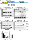
Interaction of ORFV OV20.0 with ADAR1. (A) A set of constructs expressing the wild-type form and deletions of ORFV OV20.0 were used in this study. With the Kozak sequence modification, full-length OV20.0 is preferentially expressed. (B and C) The interaction of OV20.0 with endogenous ADAR1 in a transient expression system and in ORFV-infected cells was determined by an immunoprecipitation assay (IP). Plasmids expressing FLAG-tagged wild- type OV20.0, Kozak OV20.0 (K20), short form OV20.0 (sh20), C terminus deletion OV20.0 (ΔC), and FLAG empty vector (EV, as a negative control for the IP procedure) were transfected individually into HEK 293T cells, followed by IP with an FLAG-antibody (FLAG-IP) (B). HEK 293T cells were infected with a recombinant eGFP reporter ORFV expressing FLAG-tagged OV20.0 at an MOI of 1 for 24 h, followed by FLAG-IP. Ab, antibody; M, molecular mass marker.
FIG 2
FIG 2
Cellular distribution of ADAR1 in the presence of OV20.0. The expression of endogenous ADAR1 and ADAR1 fused with mCherry was determined by an immunofluorescence assay using an ADAR1 antibody or autofluorescence, respectively. The molecular weight and integrity of mCherry (lane 1) and ADAR1-mCherry (lane 2) were determined by Western blot analysis using an mCherry antibody. (B) Colocalization of ORFV OV20.0 isoforms with ADAR1. HEK 293T cells were cotransfected with one of the eGFP-tagged OV20.0 variants (K20-eGFP, sh20-eGFP, or ΔC-eGFP) and the ADAR1-mCherry constructs. The cellular distribution of ADAR1 and OV20.0 was examined by fluorescence microscopy. White arrows indicate cytoplasmic distribution of ADAR1.
FIG 3
FIG 3
Regions of ADAR1 involved in the OV20.0 interaction. (A) The structure of ADAR1 is schematically illustrated. ADAR encodes two isoforms, namely, p150 and p110. ADAR1 contains two Z-DNA binding domains (Zα and Zβ), three RBDs (indicated as R1, R2, and R3), and the deaminase catalytic domain. Mutation of the canonical KKXXA motifs in the three RBDs to EAXXA abolished the dsRNA binding ability of ADAR1. (B) The RBDs of ADAR1 are critical for the OV20.0 interaction. Plasmids expressing HA-tagged WT ADAR1 or individual deletions of the Zβ binding domain (ΔZβ), all RBDs (ΔRBDs), or the catalytic domain (ΔCat) were cotransfected with FLAG-tagged OV20.0 (or FLAG empty vector, as a negative control for FLAG-IP) into HEK 293T cells, followed by FLAG-IP. Lane M, molecular mass marker. (C and D) OV20.0 preferentially binds R1 of ADAR1. To further explore the contribution of individual RBDs, constructs expressing deletions of one RBD (ΔR1, ΔR2, and ΔR3) were transfected along with FLAG-tagged OV20.0 into HEK 293T cells, followed by FLAG-IP. The IP experiment was performed with three independent repeats, and the relative interaction of ADAR1 with OV20.0 was then estimated. The intensity of ADAR1 variant pulldown by FLAG-IP was initially normalized to the level of β-actin. The interaction level of WT ADAR1 with OV2.0 was arbitrarily set as 1. All experiments were conducted in triplicate. *, P < 0.05.
FIG 4
FIG 4
dsRNA binding ability is not required for the interaction of OV20.0 and ADAR1. (A) Plasmids expressing HA-tagged ADAR1 (WT), dsRNA binding-deficient ADAR1 (with EAA mutations in the three RBDs; labeled EAA), and HA empty vector were transfected individually into HEK 293T cells, followed by a poly(I·C) (as a synthetic dsRNA) pulldown assay. (B) Plasmids expressing HA-tagged WT ADAR1, an EAA mutant, or the FLAG empty vector were cotransfected with FLAG-tagged OV20.0 into HEK 293T cells, followed by FLAG-IP.
FIG 5
FIG 5
OV20.0 interferes with ADAR1-dependent A-to-I-editing activity. (A) An illustration of the mCherry-based reporter system for monitoring A-to-I-editing activity. The GluR-B amber/W editing site, which contains target sequences for ADAR1-mediated RNA editing, was inserted near the C terminus of the mCherry coding region. The built-in UAG codon insertion leads to the translation of a truncated version of mCherry, while the ADAR1 enzyme likely converts A to I to yield full-length mCherry. (B) The ADAR1 enzyme edits the sequence of the reporter transcript in a dose-dependent manner. HEK 293T cells were transfected with the reporter plasmid, along with increasing amounts of HA-tagged ADAR1 (0, 0.2, 0.4, and 0.8 μg). At 24 h after transfection, A-to-I-editing activity was assessed by Western blot analysis using antibodies against HA and mCherry. (C) The effect of OV20.0 on ADAR1-mediated A-to-I editing. HEK 293T cells were cotransfected with the mCherry reporter plasmid and HA-tagged ADAR1 alone or with FLAG-tagged OV20.0 for 24 h. (D) The effect of OV20.0 on A-to-I-editing activity was measured based on the ratio of edited to the total of unedited plus edited products with increasing amounts of OV20.0 (as labeled) in the presence of ADAR1. The experiment was conducted with three independent repeats. **, P < 0.01.
FIG 6
FIG 6
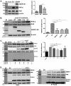
Effect of ADAR1 on PKR activation. (A) ADAR1 reduces PKR activation. HEK 293T cells were transfected with empty vector (EV) or HA-tagged ADAR1 (ADAR1), followed by activation of PKR by the transfection of 400 ng/ml poly(I·C) for 1 h. The phosphorylation (P) level of PKR was monitored by Western blot analysis. The relative PKR activation level was estimated and plotted (right panel). The PKR activation level in cells transfected with EV was arbitrarily set as 1. (B) Region required for PKR suppression. HEK 293T cells were transfected with empty vector (EV) or various ADAR1 plasmids expressing the WT protein, a deletion of the three RBD (ΔRBDs), or a deletion of the catalytic domain (ΔCat) for 24 h, and PKR phosphorylation was then induced by poly(I·C). (C) ADAR1 diminishes PKR activation in the presence of OV20.0. HEK 293T cells were cotransfected with OV20.0 (0.2 μg) and increasing amounts (0.2, 0.4, and 0.8 μg) of plasmids expressing ADAR1 overnight, followed by the induction of PKR activation. Untransfected cells under poly(I·C) stimulation served as a positive control (PC) for PKR activation; mock indicates cells without any treatment. (D) ADAR1 did not compete with PKR for the interaction with OV20.0. HEK 293T cells were cotransfected with FLAG-tagged OV20.0 (0.2 μg) with either EV or increasing amounts of the ADAR1 construct (0.1, 0.2, 0.4, and 0.8 μg), followed by FLAG-IP and immunoblotting. All the experiments were conducted in triplicate. *, P < 0.05; **, P < 0.001.
FIG 7
FIG 7
ADAR1 plays a proviral role in ORFV infection. (A) Overexpression of ADAR1 enhanced viral protein accumulation during ORFV infection. HEK 293T cells were transiently transfected with ADAR1 plasmids expressing the wild-type (WT) protein, a deletion of the three RBDs (ΔRBDs), or a deletion of the catalytic domain (ΔCat). The empty vector (EV) served as a negative control. Subsequently, the transfected cells were infected with a recombinant ORFV expressing eGFP at an MOI of 1 for 48 h, followed by fluorescence microscopy and immunoblotting. The experiment was conducted in triplicate, and the relative expression level of virus-encoded eGFP was estimated and plotted (right panel). The eGFP expression level in cells transfected with the EV was arbitrarily set as 1. (B) The proviral role of ADAR1 is correlated with its inhibitory effect on PKR activation. HEK 293T cells transiently expressing ADAR1-mCherry (ADAR1) or mCherry (EV, as a negative control) were infected with ORFV at an MOI of 1 for 48 h, followed by immunoblotting (left panel).The relative activation level of phospho-PKR (PKR-p) was normalized to the PKR basal level and plotted. The PKR activation level in cells transfected with EV was arbitrarily set as 1 (middle panel), and the yield of viral progenies was measured by standard plaque assay (right panel). (C) ORFV infection in ADAR1 knockout cells. 293T cells with endogenous (WT) ADAR1 expression or without (KO) ADAR1 expression were infected with ORFV. Expression of the viral structural protein F1L (F1 on the figure) was determined at 48 hpi by Western blot analysis (left panel). The relative expression level of F1L and yield of virus progenies were plotted (middle and right panels, respectively). All experiments were conducted in triplicate. *, P < 0.05; **, P < 0.001.

Similar articles

Cited by

References

    1. Nandi S, De UK, Chowdhury S. 2011. Current status of contagious ecthyma or orf disease in goat and sheep—a global perspective. Small Rum Res 96:73–82. doi:10.1016/j.smallrumres.2010.11.018. - DOI
    1. Fleming SB, Wise LM, Mercer AA. 2015. Molecular genetic analysis of orf virus: a poxvirus that has adapted to skin. Viruses 7:1505–1539. doi:10.3390/v7031505. - DOI - PMC - PubMed
    1. Haig DM, McInnes CJ, Thomson J, Wood A, Bunyan K, Mercer A. 1998. The Orf virus OV20.0L gene product is involved in interferon resistance and inhibits an interferon-inducible, double-stranded RNA-dependent kinase. Immunology 93:335–340. - PMC - PubMed
    1. McInnes CJ, Wood AR, Mercer AA. 1998. Orf virus encodes a homolog of the vaccinia virus interferon-resistance gene E3L. Virus Genes 17:107–115. - PubMed
    1. Tseng YY, Lin FY, Cheng SF, Tscharke D, Chulakasian S, Chou CC, Liu YF, Chang WS, Wong ML, Hsu WL. 2015. Functional analysis of the short isoform of orf virus protein OV20.0. J Virol 89:4966–4979. doi:10.1128/JVI.03714-14. - DOI - PMC - PubMed

Publication types

MeSH terms