. 2019 Mar 21;93(7):e02168-18.
doi: 10.1128/JVI.02168-18. Print 2019 Apr 1.

The Nonstructural NS1 Protein of Influenza Viruses Modulates TP53 Splicing through Host Factor CPSF4

Affiliations

The Nonstructural NS1 Protein of Influenza Viruses Modulates TP53 Splicing through Host Factor CPSF4

Julia Dubois et al. J Virol. .

Abstract

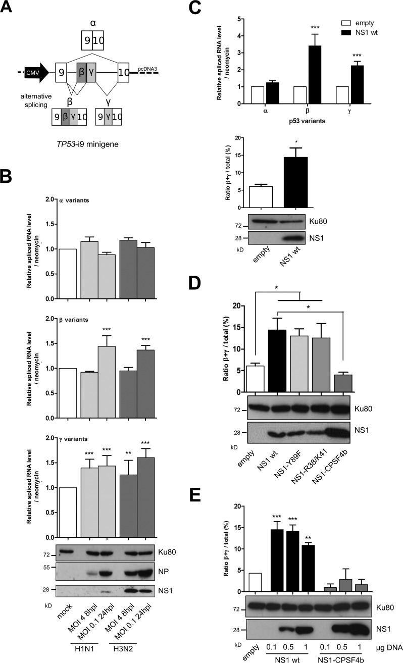
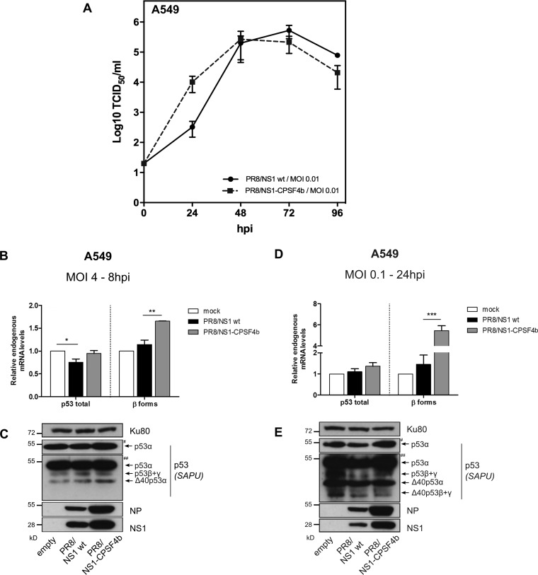
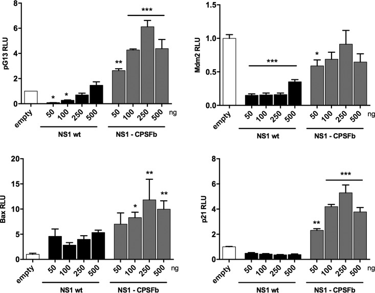
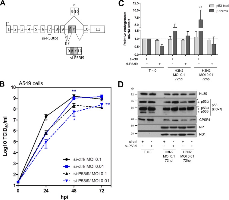
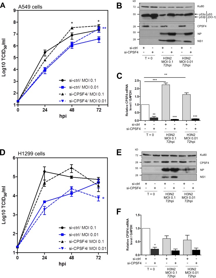
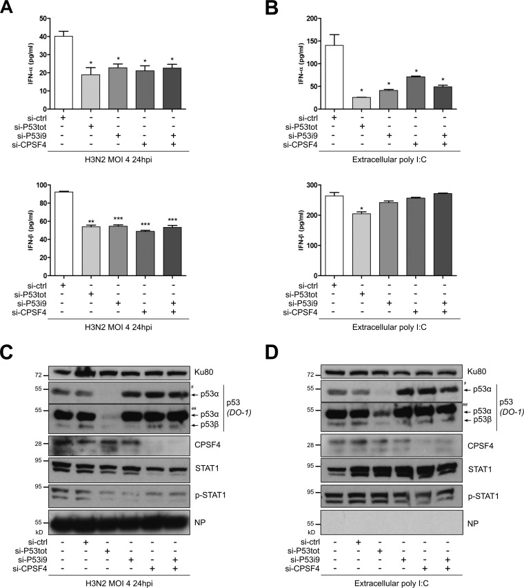
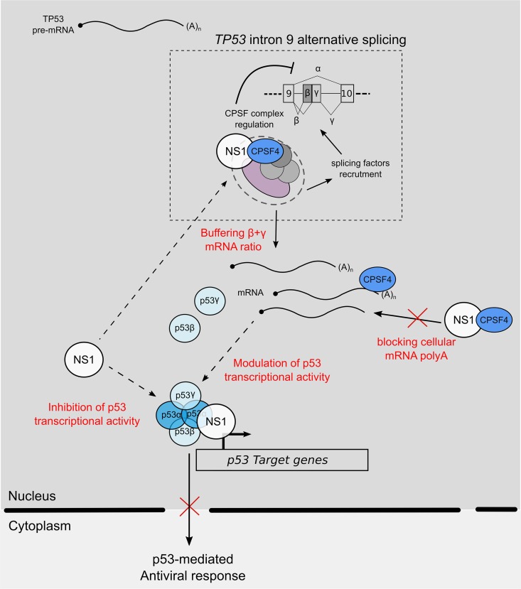
Influenza A viruses (IAV) are known to modulate and "hijack" several cellular host mechanisms, including gene splicing and RNA maturation machineries. These modulations alter host cellular responses and enable an optimal expression of viral products throughout infection. The interplay between the host protein p53 and IAV, in particular through the viral nonstructural protein NS1, has been shown to be supportive for IAV replication. However, it remains unknown whether alternatively spliced isoforms of p53, known to modulate p53 transcriptional activity, are affected by IAV infection and contribute to IAV replication. Using a TP53 minigene, which mimics intron 9 alternative splicing, we have shown here that the NS1 protein of IAV changes the expression pattern of p53 isoforms. Our results demonstrate that CPSF4 (cellular protein cleavage and polyadenylation specificity factor 4) independently and the interaction between NS1 and CPSF4 modulate the alternative splicing of TP53 transcripts, which may result in the differential activation of p53-responsive genes. Finally, we report that CPSF4 and most likely beta and gamma spliced p53 isoforms affect both viral replication and IAV-associated type I interferon secretion. All together, our data show that cellular p53 and CPSF4 factors, both interacting with viral NS1, have a crucial role during IAV replication that allows IAV to interact with and alter the expression of alternatively spliced p53 isoforms in order to regulate the cellular innate response, especially via type I interferon secretion, and perform efficient viral replication.IMPORTANCE Influenza A viruses (IAV) constitute a major public health issue, causing illness and death in high-risk populations during seasonal epidemics or pandemics. IAV are known to modulate cellular pathways to promote their replication and avoid immune restriction via the targeting of several cellular proteins. One of these proteins, p53, is a master regulator involved in a large panel of biological processes, including cell cycle arrest, apoptosis, or senescence. This "cellular gatekeeper" is also involved in the control of viral infections, and viruses have developed a wide diversity of mechanisms to modulate/hijack p53 functions to achieve an optimal replication in their hosts. Our group and others have previously shown that p53 activity is finely modulated by different multilevel mechanisms during IAV infection. Here, we characterized IAV nonstructural protein NS1 and the cellular factor CPSF4 as major partners involved in the IAV-induced modulation of the TP53 alternative splicing that was associated with a strong modulation of p53 activity and notably the p53-mediated antiviral response.

Keywords: CPSF30; antiviral response; influenza viruses; p53; splicing; virus-host interactions.


Figures

FIG 1
FIG 1
Influenza viruses and the viral NS1 protein alone modulate TP53-i9 minigene alternative splicing. (A) Schematic of the TP53-i9 minigene. Intron 9 of p53 was inserted into pCDNA3 plasmids with part of exon 9 and 10 on both sides, and transcription of mRNA corresponding to each spliced variant was under the control of the CMV promoter (47). (B) Levels of mRNA corresponding to p53 spliced variants α, β, and γ were measured in H1299 cells (p53 null) in which the TP53-i9 minigene plasmid was transfected 36 h before mock infection or infections by H1N1 or H3N2 influenza viruses. Cells lysates were harvested at 8 hpi for infection at an MOI of 4 or at 24 hpi for infection at an MOI of 0.1, and relative levels of mRNA corresponding to α, β, and γ p53 spliced variants were measured by specific RT-qPCR. The mRNA level of each variant was normalized against the neomycin resistance gene, which is also present in the TP53-i9 minigene. Viral protein expression (NP and NS1) was monitored by Western blotting. (C) The relative spliced mRNA level of p53α, p53β, and p53γ isoforms was measured in H1299 cells 48 h after cotransfection of the TP53-i9 minigene and 1 μg of an empty pCI plasmid or a plasmid containing wt NS1 (pCI NS1 wt, from influenza strain A/Moscow/10/99 [H3N2]) and was used to calculate the proportion of β+γ isoforms out of the total α, β, and γ variant p53 mRNA expression. Mean values ± standard deviations of results for at least three independent experiments are shown, and statistical tests compared each condition with its control condition using two-way analysis of variance (ANOVA) (**, P < 0.01; ***, P < 0.001). (D and E) In H1299 cells, the TP53-i9 minigene was cotransfected with 1 μg of empty pCI, pCI NS1wt (results extracted from Fig. 1C), or pCI NS1-Y89F, pCI NS1-R38A/K41A, and pCI NS1-CPSF4b mutants (D) or with increasing amounts (0.1, 0.5, or 1 μg) of pCI NS1 wt or pCI NS1-CPSF4b plasmid (E). Forty-eight hours after cotransfection, levels of α, β, and γ variant mRNA were measured (Fig. S1) and used to estimate the β+γ proportion. The efficacy of NS1 transient expression was validated by Western blotting. Mean values ± standard deviations of results from more than three independent experiments are shown, and statistical tests compared each condition with its control empty condition or NS1 wt condition using Student's t test (*, P < 0,05; **, P < 0.01; ***, P < 0.001).
FIG 2
FIG 2
IAV-regulated p53 expression is affected by NS1 CPSF4-binding mutant. (A) Comparative viral kinetics between recombinant IAV PR8/NS1 wt or PR8/NS1-CPSF4b viruses. A549 cells were infected with the two viruses at an MOI of 0.01, and supernatants were harvested at 24, 48, 72, and 96 hpi for the determination of viral titers. (B to E) A549 cells were mock infected or infected with either recombinant IAV PR8/NS1 wt or PR8/NS1-CPSF4b viruses. Cells lysates were harvested at 8 hpi (MOI of 4 [B and C]) or 24 hpi (MOI of 0.1 [D and E]). Detection of total p53 and p53β isoforms was performed by RT-qPCR and Western blotting (SAPU antibody). Viral proteins NP and NS1 were also detected. # and ## indicate short and long exposures, respectively. Mean values ± standard deviations of results of experimental duplicates are shown, and statistical tests compared each condition with its control condition using two-way ANOVA and Dunnett’s posttest (*, P < 0.05; **, P < 0.01; ***, P < 0.001).
FIG 3
FIG 3
The CPSF4-binding domain of NS1 protein plays a crucial role in the alteration of p53 isoform expression and p53 transcriptional activity. A549 cells were transfected with pG13-Luc, Mdm2-Luc, Bax-Luc or p21-Luc reporter plasmids together with increasing concentrations of an NS1-expressing plasmid (either H3N2 NS1 wt or H3N2 NS1-CPSF4b mutant). p53 transactivation activity was measured after 48 h, in triplicate in two independent experiments, and expressed in relative luciferase units (RLU) compared with the empty-plasmid condition. Mean values ± standard deviations are shown, and statistical tests compared each condition with the empty-plasmid condition using one-way ANOVA and Dunnett’s posttest (*, P < 0.05; **, P < 0.01 ***, P < 0.001).
FIG 4
FIG 4
Silencing of the TP53-i9 alternatively spliced β and γ isoforms impairs IAV replication. (A) A549 cells were treated twice with either the control nonspecific si-RNA (si-ctrl) or a specific si-RNA targeting the p53β and p53γ spliced isoforms. (B) Twenty-four hours after the last si-RNA treatment, cells were infected with H3N2 virus at an MOI of 0.1 or 0.01. Supernatants were harvested at 24-h intervals over 3 days, and the viral replication was determined by endpoint TCID50 titration in MDCK cells (measured in quadruplicate in two independent experiments). (C) Cell lysates were harvested before infection (T = 0) or at 72 hpi to quantify p53 total and p53β mRNA expression levels by RT-qPCR, normalized to actin expression. (D) Cellular and viral proteins were detected by Western blotting. # and ## indicate short and long exposures, respectively. Data represent independent experimental duplicates. Mean values ± standard deviations are shown, and statistical tests compared each condition with the si-ctrl T = 0 condition using two-way ANOVA (*, P < 0.05; **, P < 0.01).
FIG 5
FIG 5
The silencing of CPSF4 impacts IAV replication in a partially p53-dependent manner. A549 (A to C) or H1299 (D to F) cells were treated twice with either a control nonspecific siRNA (si-ctrl) or a pool of siRNAs targeting cellular CPSF4. Twenty-four hours after the last treatment, cells were infected with H3N2 at an MOI of 0.1 or 0.01. Supernatants were harvested at 24-h intervals over 3 days, and the viral kinetics in A549 cells (A) or H1299 cells (D) were determined by endpoint TCID50 titration in MDCK cells (measured in quadruplicate in two independent experiments). Cell lysates were harvested before infection (T = 0) or at 72 hpi, and cellular p53 and CPSF4, together with viral NP and NS1 proteins, were detected by Western blotting (B and E). CPSF4 mRNA expression was also measured by RT-qPCR (normalized against GAPDH expression) at T = 0 and 72 hpi (C and F). All data represent independent experimental duplicates. Mean values ± standard deviations are shown, and statistical tests compared each condition with the si-ctrl at T = 0 control using two-way ANOVA (*, P < 0.05; **, P < 0.01; ***, P < 0.001).
FIG 6
FIG 6
p53β and p53γ isoforms together with CPSF4 contribute to the p53-mediated IFN-I response to IAV infection and extracellular stresses. A549 cells were treated twice with nonspecific si-RNA (si-ctrl), an siRNA targeting all p53 forms (si-P53tot) (Fig. 4A), a specific siRNA targeting alternatively spliced p53β and p53γ isoforms (si-P53i9) (Fig. 4A), a pool of si-RNAs targeting CPSF4 (si-CPSF4), or a combination of si-P53i9 and si-CPSF4. Twenty-four hours later, cells were infected with H3N2 at an MOI of 4 (A and C) or extracellular poly(I·C) was added to induce a stress through TLR3 activation (B and D). (A and B) Supernatants were harvested 24 h after treatment, and IFN-α and IFN-β levels were quantified. (C) Cell lysates were also collected to monitor infection, si-RNA efficiency, and type I IFN response via STAT1 phosphorylation by Western blotting. # and ## indicate short and long exposures, respectively, with DO-1 antibody. Mean values ± standard deviations of results of experimental duplicates are shown, and the two-way ANOVA test compared each condition with the si-ctrl condition (*, P < 0.05; **, P < 0.01; ***, P < 0.001).
FIG 7
FIG 7
Working model of interplay between IAV NS1 protein, cellular factor CPSF4, and TP53 splicing. During IAV infection, IAV NS1 inhibits p53 transcriptional activity via its interaction with p53 but also via the modulation of TP53 splicing by “buffering” the function of CPSF4 in mRNA maturation and splicing. As a result, the spliced p53 isoform modulation of p53 transcriptional activity, and notably p53-mediated antiviral responses, coupled to the cellular impact of CPSF4 blockade positively influences viral production. When NS1 is mutated, preventing its binding to CPSF4, this regulation loop is impaired, and the antiviral response is increased, limiting viral production.

Similar articles

Cited by

References

    1. Paules C, Subbarao K. 2017. Influenza. Lancet 390:697–708. - PubMed
    1. Neumann G, Kawaoka Y. 2011. The first influenza pandemic of the new millennium. Influenza Other Respir Viruses 5:157–166. doi:10.1111/j.1750-2659.2011.00231.x. - DOI - PMC - PubMed
    1. Palese PSM. 2013. Orthomyxoviridae, p 1151–1185. In Knipe DM, Howley P (ed), Fields virology, 6th ed Lippincott Williams & Wilkins, Philadelphia, PA.
    1. Watanabe T, Watanabe S, Kawaoka Y. 2010. Cellular networks involved in the influenza virus life cycle. Cell Host Microbe 7:427–439. doi:10.1016/j.chom.2010.05.008. - DOI - PMC - PubMed
    1. Dubois J, Terrier O, Rosa-Calatrava M. 2014. Influenza viruses and mRNA splicing: doing more with less. mBio 5:e00070. doi:10.1128/mBio.00070-14. - DOI - PMC - PubMed

Publication types

MeSH terms

Substances