Review
. 2019 Apr;15(4):220-239.
doi: 10.1038/s41581-018-0103-6.

Epigenetic regulation in AKI and kidney repair: mechanisms and therapeutic implications

Affiliations
Review

Epigenetic regulation in AKI and kidney repair: mechanisms and therapeutic implications

Chunyuan Guo et al. Nat Rev Nephrol. 2019 Apr.

Abstract

Acute kidney injury (AKI) is a major public health concern associated with high morbidity and mortality. Despite decades of research, the pathogenesis of AKI remains incompletely understood and effective therapies are lacking. An increasing body of evidence suggests a role for epigenetic regulation in the process of AKI and kidney repair, involving remarkable changes in histone modifications, DNA methylation and the expression of various non-coding RNAs. For instance, increases in levels of histone acetylation seem to protect kidneys from AKI and promote kidney repair. AKI is also associated with changes in genome-wide and gene-specific DNA methylation; however, the role and regulation of DNA methylation in kidney injury and repair remains largely elusive. MicroRNAs have been studied quite extensively in AKI, and a plethora of specific microRNAs have been implicated in the pathogenesis of AKI. Emerging research suggests potential for microRNAs as novel diagnostic biomarkers of AKI. Further investigation into these epigenetic mechanisms will not only generate novel insights into the mechanisms of AKI and kidney repair but also might lead to new strategies for the diagnosis and therapy of this disease.


Figures
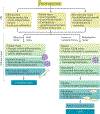
Fig. 1 |
Fig. 1 |. Pathophysiology of AKI and repair.
The pathophysiology of acute kidney injury (AKI) is very complex, involving interplay between tubular, microvascular and inflammatory factors. Acute injury insults typically induce the injury and death of tubular epithelial cells, injury and activation of endothelial cells and leukocyte infiltration, culminating in renal dysfunction. In the presence of mild injury, adaptive repair mechanisms can restore epithelial integrity, suppress the immune response and re-establish healthy vasculature. By contrast, severe or persistent injury induces maladaptive repair. Tubular cells may undergo G2/M cell cycle arrest, senescence and apoptosis or necrosis, leading to the release of profibrotic and pro-inflammatory factors. Tubular atrophy and degeneration, together with sustained inflammation and microvascular loss, result in renal interstitial fibrosis, characterized by the proliferation and activation of fibroblasts and deposition of extracellular matrix (ECM), ultimately leading to chronic kidney disease (CKD).
Fig. 2 |
Fig. 2 |. Mechanisms and consequences of histone modifications.
a | Histone modifications (acetylation, methylation and phosphorylation) are catalysed by specific enzymes known as epigenetic writers, recognized by epigenetic readers and removed by epigenetic erasers. b | The major sites of histone acetylation, methylation and phosphorylation. Generally, histone acetylation is associated with permissive transcription, and histone methylation is associated with either active (H3 lysine 4 (H3K4), H3K36 and H3K79) or repressive (H3K9, H3K27 and H4K20) transcription. Phosphorylation of histones (for example, phosphorylation of H3 at serine 10 (H3S10ph), threonine 11 (H3T11ph), serine 28 (H3S28ph) and tyrosine 41 (H3Y41ph) and phosphorylation of H2B at serine 32 (H2BS32ph)) is often associated with transcriptional activation; however, histone phosphorylation (for example, of H2A at serine 1 (H2AS1ph), phosphorylation of H2B at tyrosine 37 (H2BY37ph) and phosphorylation of H4 at serine 1 (H4S1ph)) can be associated with transcriptional repression. HATs, histone acetyltransferases; HDACs, histone deacetylases; HDMs, histone demethylases; HMTs, histone methyltransferases.
Fig. 3 |
Fig. 3 |. Mechanisms of DNA methylation and demethylation.
a | DNA methylation involves the covalent addition of a methyl group (CH3) to cytosine by DNA methyltransferases (DNMTs). Initial DNA methylation patterns are established by DNMT3a and DNMT3b. When DNA replication occurs, DNMT1 faithfully copies the DNA methylation patterns from parental strands to daughter strands. DNA methylation can inhibit gene expression directly by preventing transcription factor binding (1) or indirectly through the actions of DNA methylation readers, such as methyl-CpG-binding domain (MBD) proteins, which can either inhibit the binding of transcription factors (2) or recruit transcription repressors (3), leading to gene suppression. b | The process of DNA demethylation can be passive or active. Passive demethylation occurs in the absence of functional DNMT1 during cell division, as newly synthesized DNA strands are not able to be methylated. Methyl groups can also be actively removed from DNA by an enzymatic replacement mechanism called active demethylation. On one hand, 5-methylcytosine (5mC; a methylated form of cytosine) can be deaminated to thymine by activation-induced cytidine deaminase (AID), which generates a mismatch between thymine and guanine bases. 5mC can also be oxidized by ten-eleven translocation (TET) proteins to 5-hydroxymethylcytosine (5hmC), which can be further oxidized to 5-formylcytosine (5fC) and 5-carboxylcytosine (5caC) or deaminated to 5-hydroxymethyl-uracil (5hmU). Finally, these intermediates are replaced by cytosine through thymine DNA glycosylase (TDG) and base excision repair (BER). SAH, S-adenosyl-L-homocysteine; SAM, S‑adenosyl-L-methionine.
Fig. 4 |
Fig. 4 |. Mechanisms of gene regulation by non-coding RNAs.
a | MicroRNA biogenesis requires initial transcription of a microRNA gene by RNA polymerase II into a large primary transcript that contains multiple hairpin loop structures, known as a primary microRNA (pri-microRNA). This transcript is processed by the ribonuclease, Drosha, exported from the nucleus and processed by another enzyme, called Dicer, to produce a microRNA–microRNA duplex in the cytoplasm consisting of a guide strand and a passenger strand. This complex is unwound and the mature microRNA (the guide strand) is incorporated into the ribonucleoprotein complex, RISC (RNA-induced silencing complex), which is guided to target genes, leading to post-transcriptional silencing either by mRNA degradation or most commonly by translational repression. b | Long non-coding RNAs (lncRNAs) can regulate gene expression at both transcriptional (1–4) and post-transcriptional levels (5–7). At a transcriptional level, lncRNAs can (1) recruit transcription activators or repressors to the target gene, resulting in gene activation or suppression, respectively; (2) act as decoy factors by binding and sequestering transcription factors and other proteins or regulatory RNAs away from chromatin, thereby inhibiting gene transcription; (3) guide chromatin-modifying enzymes to specific gene targets and then modulate chromatin states either in cis or trans, leading to positive or negative regulation of gene expression; and (4) serve as a structural scaffold to recruit multiple proteins or RNAs to form transcription regulatory complexes, which can positively or negatively regulate gene expression. lncRNAs can also regulate gene expression at a post-transcriptional level by (5) binding to splicing factors to regulate RNA splicing, (6) directing RNA-binding proteins to the target mRNAs to either promote or inhibit protein translation or (7) serving as a molecular sponge that binds and titrates microRNAs away from their targets.

References

    1. Lameire NH et al. Acute kidney injury: an increasing global concern. Lancet 382, 170–179 (2013). - PubMed
    1. Mehta RL et al. Recognition and management of acute kidney injury in the International Society of Nephrology 0by25 Global Snapshot: a multinational cross-sectional study. Lancet 387, 2017–2025 (2016). - PubMed
    1. Mehta RL et al. International Society of Nephrology’s 0by25 initiative for acute kidney injury (zero preventable deaths by 2025): a human rights case for nephrology. Lancet 385, 2616–2643 (2015). - PubMed
    1. Linkermann A. et al. Regulated cell death in AKI. J. Am. Soc. Nephrol 25, 2689–2701 (2014). - PMC - PubMed
    1. Agarwal A. et al. Cellular and molecular mechanisms of AKI. J. Am. Soc. Nephrol 27, 1288–1299 (2016). - PMC - PubMed

Publication types