

Lung lobe torsion in 35 dogs and 4 cats
- PMID: 30651652
- PMCID: PMC6294018
Lung lobe torsion in 35 dogs and 4 cats
Abstract
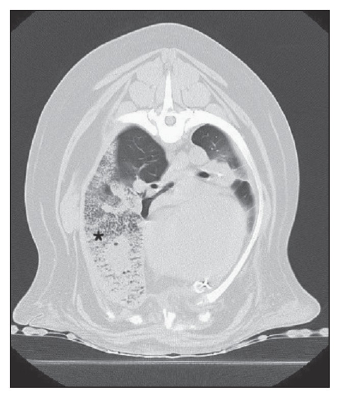
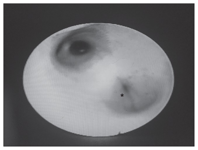
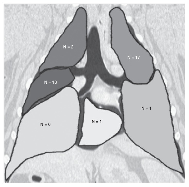
The purpose of this study was to assess survival to discharge of animals with surgical or postmortem confirmation of a lung lobe torsion (LLT) as well as to evaluate pre-operative effusion, lung lobe affected, and patient size as prognostic indicators. Medical records search identified 35 dogs and 4 cats with a confirmed diagnosis including 17 small-breed dogs, 18 large-breed dogs, 3 domestic shorthair cats, and 1 minskin cat. Lobes affected included right middle (n = 18), left cranial (n = 18), right cranial (n = 2), left caudal (n = 1), and accessory (n = 1). Two animals died before surgery; the remaining 37 animals underwent thoracotomy. All treated small-breed dogs and cats survived; 12/18 large-breed dogs survived, with an overall survival to discharge of 87%. Pre-operative pleural effusion and affected lung lobe did not affect survival to discharge in this population. Small dogs and cats with LLT appear to have an excellent survival to discharge following thoracotomy and the survival is good in larger dogs.
Torsion de lobe pulmonaire chez 35 chiens et 4 chats. Le but de cette étude est d’évaluer le taux de survie jusqu’à la sortie d’hôpital, chez des animaux ayant eu confirmation chirurgicale ou post-mortem de torsion de lobe pulmonaire (TLP), ainsi que d’évaluer la présence d’épanchement pleural, le lobe pulmonaire affecté et la taille des patients en tant que facteurs pronostics. Les dossiers médicaux de 35 chiens et 4 chats avec un diagnostic de TLP confirmé furent identifiés, comprenant 17 chiens de petite race, 18 chiens de grande race, 3 chats Européens et 1 Minskin. Les lobes pulmonaires affectés étaient le lobe moyen droit (n = 18), le lobe cranial gauche (n = 18), le lobe cranial droit (n = 2), le lobe caudal gauche (n = 1) et le lobe accessoire (n = 1). Deux animaux décédèrent avant chirurgie, et une thoracotomie fut pratiquées chez les 37 animaux restant. Tous les chiens de petites races et les chats survécurent à leur chirurgie; 12/18 des chiens de grande race survécurent, et le taux de survie global était de 87 %. La présence d’épanchement pleural et le lobe pulmonaire affecté n’avaient pas d’influence sur la probabilité de survie dans cette population. Le taux de survie au congé hospitalier semble excellent chez les chiens de petite taille et les chats diagnostiqués avec une torsion de lobe pulmonaire et il est bon pour les chiens de grande taille.(Traduit par les auteurs).
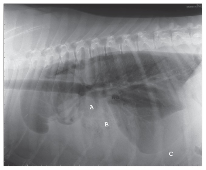
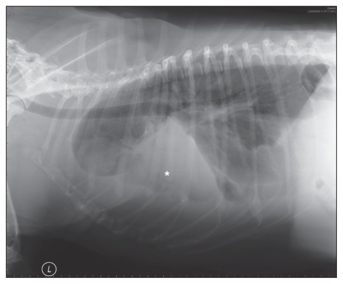
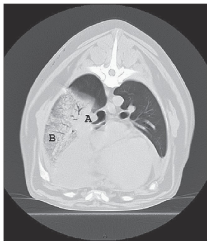
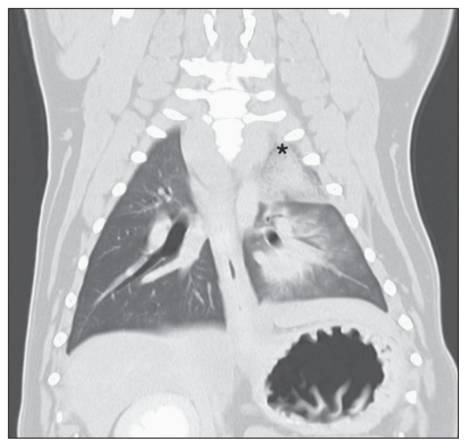
Figures
Similar articles
-
Clinical characteristics and long-term outcome of lung lobe torsions in cats: a review of 10 cases (2000-2021).J Feline Med Surg. 2022 Oct;24(10):1072-1080. doi: 10.1177/1098612X211054816. Epub 2021 Nov 1. J Feline Med Surg. 2022. PMID: 34719995 Free PMC article. Review.
-
Lung lobe torsion.Compend Contin Educ Vet. 2011 Apr;33(4):E4. Compend Contin Educ Vet. 2011. PMID: 21870341 Review.
-
Radiographic diagnosis of lung lobe torsion.Vet Radiol Ultrasound. 2005 Nov-Dec;46(6):478-84. doi: 10.1111/j.1740-8261.2005.00087.x. Vet Radiol Ultrasound. 2005. PMID: 16396263
-
Lung lobe torsion in association with a chronic diaphragmatic hernia and haemorrhagic pleural effusion in a cat.J Feline Med Surg. 2012 Mar;14(3):219-23. doi: 10.1177/1098612X12439270. J Feline Med Surg. 2012. PMID: 22370865 Free PMC article.
-
Lung lobe torsion in dogs: 22 cases (1981-1999).J Am Vet Med Assoc. 2000 Oct 1;217(7):1041-4. doi: 10.2460/javma.2000.217.1041. J Am Vet Med Assoc. 2000. PMID: 11019713
Cited by
-
Clinical characteristics and long-term outcome of lung lobe torsions in cats: a review of 10 cases (2000-2021).J Feline Med Surg. 2022 Oct;24(10):1072-1080. doi: 10.1177/1098612X211054816. Epub 2021 Nov 1. J Feline Med Surg. 2022. PMID: 34719995 Free PMC article. Review.
-
Bilateral lung lobe torsions in a cat.Can Vet J. 2020 Feb;61(2):161-165. Can Vet J. 2020. PMID: 32020935 Free PMC article.
-
Therapy of Pyothorax in Cats via Small-Bore Thoracostomy Tube in Terms of Efficacy, Complications and Outcomes.Animals (Basel). 2022 Jan 3;12(1):107. doi: 10.3390/ani12010107. Animals (Basel). 2022. PMID: 35011213 Free PMC article.
References
-
- d’Anjou MA, Tidwell AS, Hecht S. Radiographic diagnosis of lung lobe torsion. Vet Radiol Ultrasound. 2005;46:478–484. - PubMed
-
- Neath PR, Brockman DJ, King LG. Lung lobe torsion in dogs: 22 cases (1981–1999) J Am Vet Med Assoc. 2000;217:1041–1044. - PubMed
-
- Murphy KA, Brisson BA. Evaluation of lung lobe torsion in Pugs: 7 cases (1991–2004) J Am Vet Med Assoc. 2006;228:86–90. - PubMed
-
- Gicking JC, Aumann M. Lung lobe torsion. Compend Contin Educ Vet. 2011;44:E1–E4. - PubMed
-
- Monnet E. Lungs. In: Tobias KM, Johnston SA, editors. Veterinary Surgery Small Animal. St Louis, Missouri: Saunders Elsevier; 2012. pp. 1752–1768.
MeSH terms
LinkOut - more resources
Full Text Sources
Medical
Miscellaneous