. 2018 Sep-Dec;22(3):448.
doi: 10.4103/jomfp.JOMFP_33_18.

Demystifying the nature of hard tissues in odontogenic tumors using Modified Gallego's stain: A preliminary study

Affiliations

Demystifying the nature of hard tissues in odontogenic tumors using Modified Gallego's stain: A preliminary study

Syeda Neelam Afroze et al. J Oral Maxillofac Pathol. 2018 Sep-Dec.

Abstract

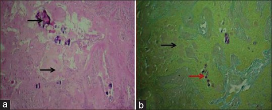
Introduction: Histological stains are dyes that bind to a variety of tissues. Modified Gallego's (MG) stain is a modification of Lille's stain that can be used as a differential stain for identification of hard tissues in oral pathological lesions.

Objectives: The objective of this study was to identify the presence of hard tissues such as enamel, dentin and cementum in normal extracted teeth and odontogenic tumors using MG stain and to compare the efficacy of MG stain with hematoxylin and eosin (H&E) stain.

Methods: A total of fifty samples, twenty decalcified sections of teeth and thirty cases of odontogenic tumors, were included in the present study. Two sections were cut from the above cases and stained with H&E stain and MG stain, respectively, and assessed for the nature of hard tissue.

Results: In H&E staining, enamel, dentine, cementum and bone stained pink. Whereas, in MG stain, enamel stained pink, dentin and bone stained green, while cementum stained red. The shade of color differs with the degree of mineralization of the hard tissues in MG stain.

Conclusion: MG stain can be used as a differential stain for different hard-tissue structures when compared to routine H and E staining.

Keywords: Hematoxylin and eosin stain; Modified Gallego's stain; odontogenic tumors.


Conflict of interest statement

There are no conflicts of interest.

Figures

Figure 1
Figure 1
Photomicrograph of decalcified section in hematoxylin and eosin and Modified Gallego's stains. (A) Decalcified section of teeth in hematoxylin and eosin ([a] Dentin – pink, [b] cementum – pink). (B) Decalcified section of teeth in Modified Gallego's stain ([a] Dentin – green, [b] Cementum – red). (C) Decalcified section of bone in hematoxylin and eosin. (D) Decalcified section of bone in Modified Gallego's stain (bone – green)
Figure 2
Figure 2
Photomicrograph of odontome: (a) Hematoxylin and eosin stain showing pink-colored deposits. (b) Modified Gallego's stain showing dentin/bone-like material in green (black arrow) and cementum-like material in red color (red arrow)
Figure 3
Figure 3
Photomicrograph of dentinogenic ghost cell tumor: (a) Hematoxylin and eosin stain showing pink-colored deposits. (b) Modified Gallego's stain showing dentin/bone-like material in green (black arrow)
Figure 4
Figure 4
Photomicrograph of ameloblastic fibro dentinoma: (a) Hematoxylin and eosin stain showing pink-colored deposits. (b) Modified Gallego's stain showing dentin/bone-like material in green (black arrow)
Figure 5
Figure 5
Photomicrograph of calcifying epithelial odontogenic tumor: (a) Hematoxylin and eosin stain showing pink-colored deposits. (b) Modified Gallego's stain showing amyloid-like material in pink (black arrow)
Figure 6
Figure 6
Photomicrograph of odontoameloblastoma: (a) Hematoxylin and eosin stain showing pink-colored deposits. (b) Modified Gallego's stain showing dentin/bone-like material in green color (black arrow) and enamel-like material in pink (red arrow)
Figure 7
Figure 7
Photomicrograph of adenomatoid odontogenic tumor: (a) Hematoxylin and eosin stain showing pink-colored deposits. (b) Modified Gallego's stain showing enamel-like material in pink (black arrow)

References

    1. Alturkistani HA, Tashkandi FM, Mohammedsaleh ZM. Histological stains: A Literature review and case study. Glob J Health Sci. 2015;8:72–9. - PMC - PubMed
    1. Culling CF. Histopathological and Histochemical Techniques. 3rd ed. London: Butterworth; 1974. pp. 63-72–420-4.
    1. Bancroft JD, Gamble M. Theory and Practice of Histological Technique. 5th ed. New York: Churchill Livingstone; 1996. 139 pp.
    1. Ramulu S, Kale AD, Hallikerimath S, Kotrashetti V. Comparing modified Papanicolaou stain with Ayoub-Shklar and haematoxylin-eosin stain for demonstration of keratin in paraffin embedded tissue sections. J Oral Maxillofac Pathol. 2013;17:23–30. - PMC - PubMed
    1. Tamgadge SA, Tamgadge A, Srivastava C, Satheesan E, Bhalerao S. Modified Gallego's stain as differential stain for oral hard tissues in oral pathology: A preliminary report. Int J Oral Maxillofac Pathol. 2014;5:2–6.