. 2019 Jan;17(1):883-887.
doi: 10.3892/etm.2018.7002. Epub 2018 Nov 21.

Clinical observation of orbital IgG4-related diseases

Affiliations

Clinical observation of orbital IgG4-related diseases

Na Wu et al. Exp Ther Med. 2019 Jan.

Abstract

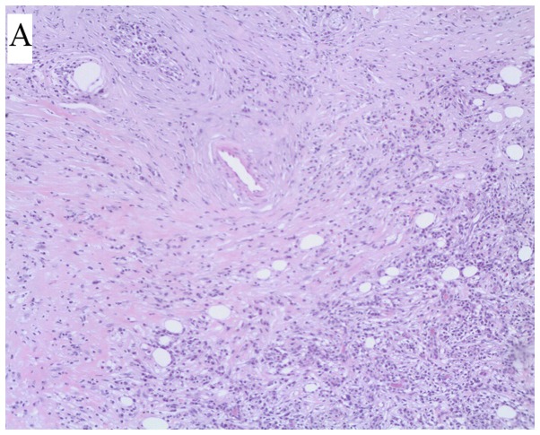
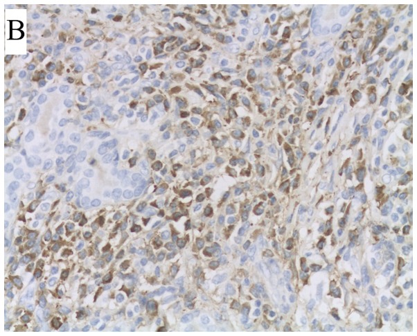
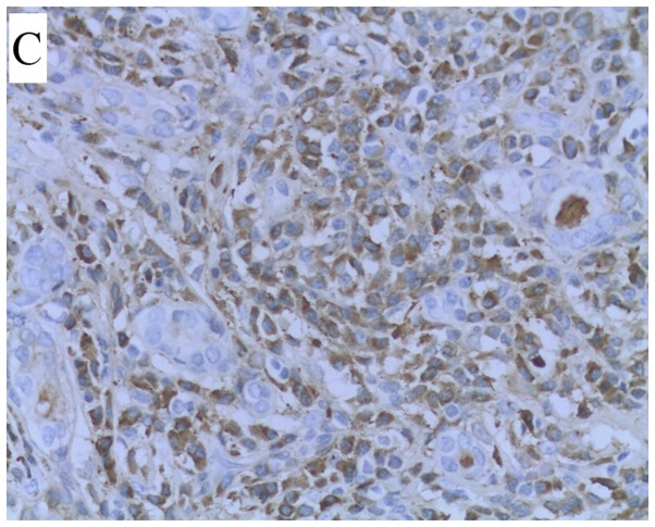
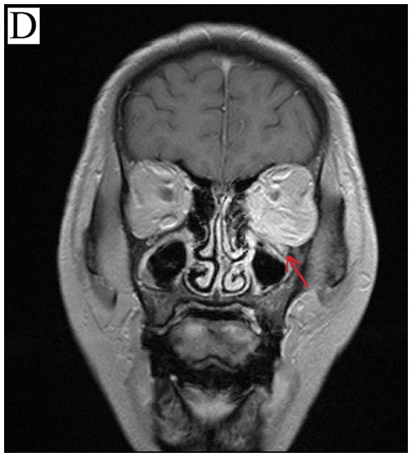
The aim of the present study was to observe the histopathological changes of immunoglobulin G4-related orbital diseases (IgG4-RODs), summarize the clinical manifestations and imaging features of the IgG4-RODs of the eyelids and explore the early diagnosis of IgG4-RODs. Between June 2011 and May 2015, 23 patients with non-specific orbital inflammation in the Department of Ophthalmology at the First Central Hospital of Tianjin were recruited. The serum IgG4 titer in 9 patients ranged from 4.58 to 46.70 g/l (reference value, 0.03-2.01 g/l), with an average value of 21.93±2.18 g/l. Notably, the degree of increase in the 9 patients with IgG4-RODs was different, but all were >1.35 g/l. A total of 6 cases of infraorbital nerve thickening were observed. In addition, there were 3 cases of extraocular muscle thickening and 1 patient with IgG4-ROD had an orbital tissue lesion extending along the inferior temporal septum to the left pterygopalatine fossa, with left sacral fissure widening and involvement of the left maxillary sinus. The study revealed that the thickening of the inferior orbital nerve may be a characteristic of IgG4-ROD. Therefore, on the basis of biopsy and serological examination in the clinic, early diagnosis can be combined with imaging examination, clinical manifestation and laboratory examination, so as to reduce misdiagnosis and missed diagnosis.

Keywords: IgG4-related eye disease; differential diagnosis; immunoglobulin G; immunohistochemistry; orbital disease.


Figures

Figure 1.
Figure 1.
Evaluation of a patient with IgG4-ROD following orbital lesion resection. (A) A large amount of lymphocyte and cytoplasmic infiltration and cystia in the lesions were observed via hematoxylin and eosin staining (magnification, ×50). (B) A large number of IgG+ cells were indicated in the diseased tissue using EnVision (magnification, ×400). (C) A large number of IgG4+ cells were demonstrated in the diseased tissue (magnification, ×400). (D) Nerve enlargement of left eye socket.
Figure 1.
Figure 1.
Evaluation of a patient with IgG4-ROD following orbital lesion resection. (A) A large amount of lymphocyte and cytoplasmic infiltration and cystia in the lesions were observed via hematoxylin and eosin staining (magnification, ×50). (B) A large number of IgG+ cells were indicated in the diseased tissue using EnVision (magnification, ×400). (C) A large number of IgG4+ cells were demonstrated in the diseased tissue (magnification, ×400). (D) Nerve enlargement of left eye socket.

References

    1. McNab AA, McKelvie P. IgG4-related ophthalmic disease. Part I: Background and pathology. Ophthal Plast Reconstr Surg. 2015;31:83–88. doi: 10.1097/IOP.0000000000000363. - DOI - PubMed
    1. Patnana M, Sevrukov AB, Elsayes KM, Viswanathan C, Lubner M, Menias CO. Inflammatory pseudotumor: The great mimicker. AJR Am J Roentgenol. 2012;198:W217–W227. doi: 10.2214/AJR.11.7288. - DOI - PubMed
    1. Sato Y, Ohshima K, Ichimura K, Sato M, Yamadori I, Tanaka T, Takata K, Morito T, Kondo E, Yoshino T. Ocular adnexal IgG4-related disease has uniform clinicopathology. Pathol Int. 2008;58:465–470. doi: 10.1111/j.1440-1827.2008.02257.x. - DOI - PubMed
    1. Jin R, Zhao P, Ma X, Ma J, Wu Y, Yang X, Zhang J, Zhong R, Zeng Y. Quantification of Epstein-Barr virus DNA in patients with idiopathic orbital inflammatory pseudotumor. PLoS One. 2013;8:e50812. doi: 10.1371/journal.pone.0050812. - DOI - PMC - PubMed
    1. Mombaerts I, Goldschmeding R, Schlingemann RO, Koornneef L. What is orbital pseudotumor? Surv Ophthalmol. 1996;41:66–78. doi: 10.1016/S0039-6257(97)81996-0. - DOI - PubMed