

Review
doi: 10.3941/jrcr.v12i5.3234.
eCollection 2018 May.
"Mixed" trauma to the carotid artery in a mixed martial arts injury - A case report and review of the literature
Affiliations
- PMID: 30651908
- PMCID: PMC6310208
- DOI: 10.3941/jrcr.v12i5.3234
Item in Clipboard
Review
"Mixed" trauma to the carotid artery in a mixed martial arts injury - A case report and review of the literature
J Radiol Case Rep.
.
Abstract
We present the case of a mixed martial arts (MMA) cage fighter who presented to the emergency department with a right sided common carotid artery pseudoaneurysm as a result of a neck trauma at an MMA event. We discuss the management of blunt force neck trauma, differential diagnosis, imaging findings and review the literature on blunt cerebrovascular injury following blunt force injury to the neck.
Keywords: Blunt cerebrovascular trauma; Carotid arterial injury; pseudoaneurysm; thyroid horn fracture; trauma.
Figures
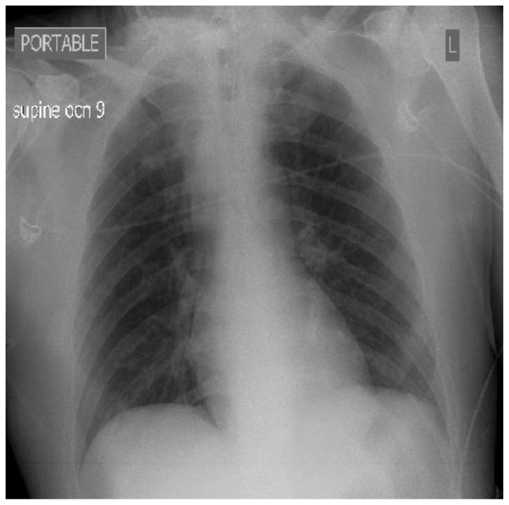
36 year old male imaged following right sided blunt neck trauma resulting in a right carotid artery pseudoaneurysm. Portable supine plain film chest x ray demonstrating slight widening of the mediastinum. Findings: Slight mediastinal widening. Technique: Portable Supine Plain film chest X-ray, Fujifilm FDR Go system, 80 kVp, 30
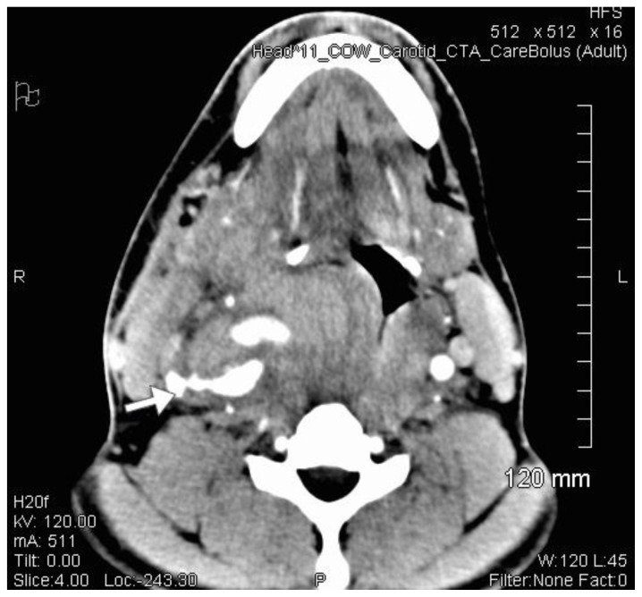
36 year old male imaged following blunt force trauma to right side of the neck. Axial CT Angiogram of the Carotid arteries demonstrating a right sided Carotid artery pseudoaneurysm. Findings: Right sided carotid artery pseudoaneurysm. Technique: Siemens SOMATOM 64 slice CT scanner, Axial CT Angiogram, 511mA, Contrast: 80mls Omnipaque 300 at 4.5mls/second, 120kV, 1mm slice thickness
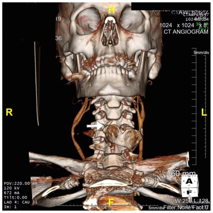
36 year old male imaged following blunt force trauma to right side of the neck resulting in a right carotid artery pseudoaneurysm. Findings: Fracture of the right superior horn of the thyroid cartilage (white arrow), with lateral bowing of the common carotid artery proximal to the carotid bifurcation suggesting extramural compression from pseudoaneurysm seen on CTA imaging. Technique: Siemens SOMATOM 64 slice CT scanner, Coronal 3D CT reformat, 672mA, 120KV, 1mm slice thickness)
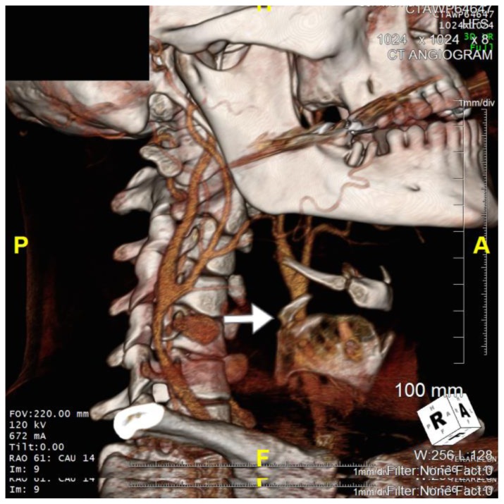
36 year old male imaged following blunt force trauma to right side of the neck resulting in a right carotid artery pseudoaneurysm. Findings: Fracture of the right superior horn of the thyroid cartilage (white arrow), with lateral bowing of the common carotid artery proximal to the carotid bifurcation suggesting extramural compression from the pseudoaneurysm seen on CTA imaging Technique: Siemens SOMATOM 64 slice CT scanner, Right Anterooblique (RAO) view, Axial 3D CT reformat, 672mA, 120kV, 1mm slice thickness, Tilt 0 degrees, RAO 61 degrees, CAU 14 degrees)
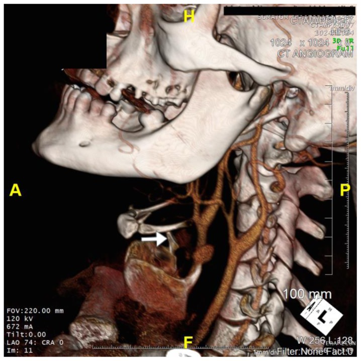
36 year old male imaged following blunt force trauma to right side of the neck resulting in a right carotid artery pseudoaneurysm. Findings: Fracture of the right superior horn of the thyroid cartilage (white arrow), with lateral bowing of the common carotid artery proximal to the carotid bifurcation suggesting extramural compression from the pseudoaneurysm seen on CTA imaging Technique: Siemens SOMATOM 64 slice CT scanner, Axial Left Anterooblique (LAO) projection 3D CT reformat, 672mA, 120kV, 1mm slice thickness, Tilt 0 degrees, LAO 74 degrees, Cranial 0 degrees)
References
-
- Welling R, Kakkasseril J, Peschiera J. Management of blunt injury to the internal carotid artery. J Trauma. 1987;27:1221–6. - PubMed
-
- Schneidereit NP, Simons R, Nicolaou S, et al. Utility of screening for blunt vascular neck injuries with computed tomographic angiography. J Trauma. 2006;60(1):209–15. 5. - PubMed
-
- Sliker CW. Blunt cerebrovascular injuries: imaging with multidetector CT angiography. Radiographics. 2008;28(6):1689–708. - PubMed
-
- Fox C, Gillespie D, Weber M, et al. Delayed evaluation of combat related penetrating neck trauma. J Vasc Surg. 2006;44:86–92. - PubMed
-
- Garg Karan, et al. Presentation and management of carotid artery aneurysms and pseudo aneurysms. J Vasc Surg. 2012;55(6):1618–1622. - PubMed
Publication types
MeSH terms
LinkOut - more resources
Full Text Sources