Case Reports
. 2018 Sep 30;12(9):21-30.
doi: 10.3941/jrcr.v12i9.3390. eCollection 2018 Sep.

Imaging features of uterine and ovarian fibromatosis in Nevoid Basal Cell Carcinoma Syndrome

Affiliations
Case Reports

Imaging features of uterine and ovarian fibromatosis in Nevoid Basal Cell Carcinoma Syndrome

Ambra Cassar Scalia et al. J Radiol Case Rep. .

Abstract

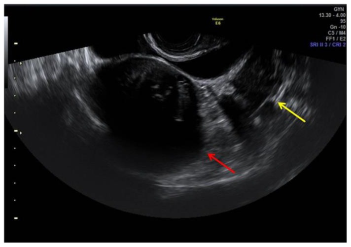
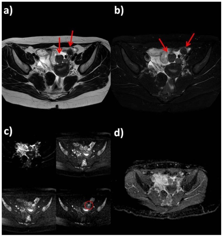
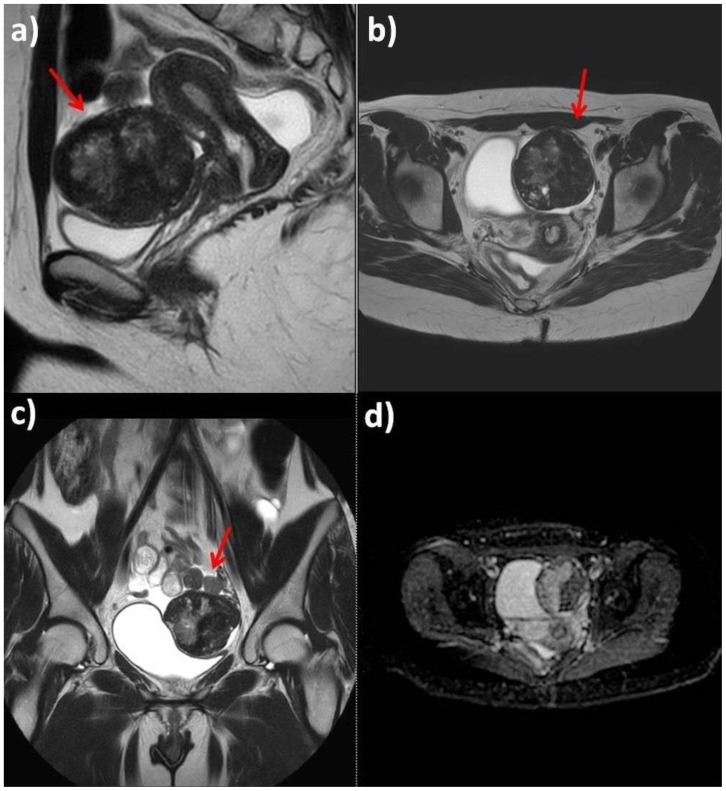
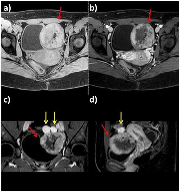
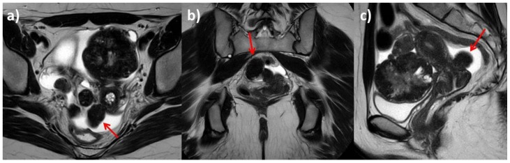
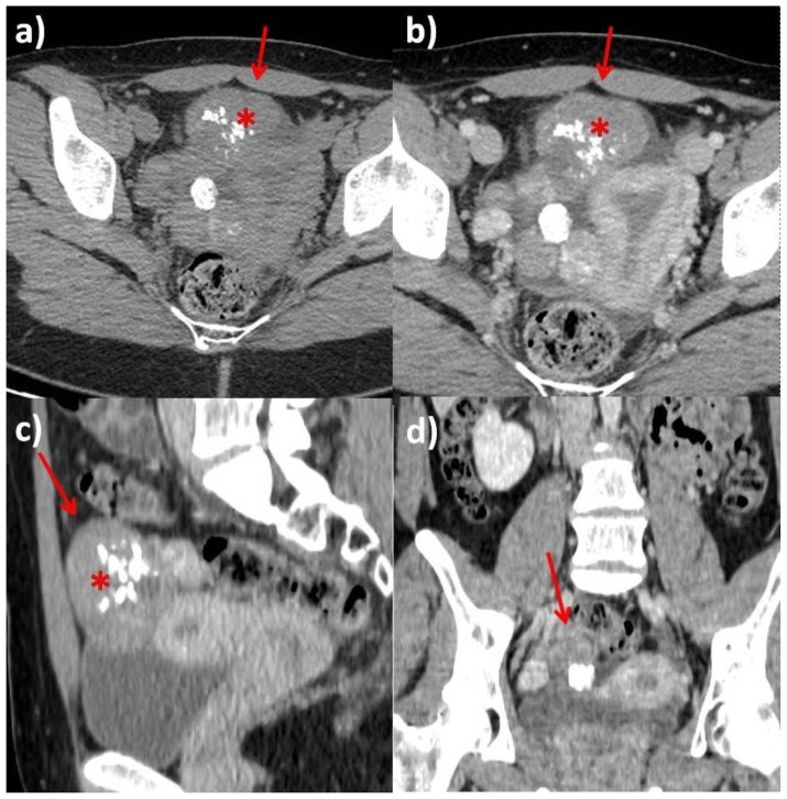
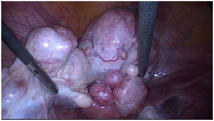
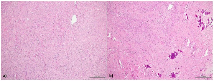
Gorlin-Goltz Syndrome also known as Nevoid Basal Cell Carcinoma Syndrome is an autosomal dominant multisystem disorder. It is characterized by basal cell carcinomas, odontogenic keratocysts, skeletal abnormalities and in a minority of female patients bilateral calcified ovarian fibromas. It is challenging to radiologically assess ovarian fibromas as they have similar imaging patterns to some malignant ovarian lesions. However, it is vitally important to differentiate between benign and malignant lesions to determine patients' suitability for fertility-sparing surgery. This report describes a case of a 25 year-old patient with Gorlin-Goltz Syndrome and bilateral ovarian fibromas.

Keywords: Gorlin-Goltz syndrome; MRI; NBCCS; ovarian fibromas; uterine fibromas.


Figures

Figure 1
Figure 1
25-year-old woman with multiple ovarian and uterine fibromas in Nevoid Basal Cell Carcinoma Syndrome. Findings: ultrasound imaging highlights the presence of heterogenous hypoechoic uterine round-shaped mass measuring 65 × 62 × 59 mm adherent to the anterior uterine wall (red arrow). Left ovary was appreciable with difficulty due to the presence of a rounded hypoechoic mass measuring 18 × 15 mm (yellow arrow: an ovarian mass). Ovarian follicles were not depictable bilaterally. Technique: General Electric Voluson E6 (transrectal transducer, 4–9 MHz).
Figure 2
Figure 2
25-year-old woman with multiple ovarian and uterine fibromas in Nevoid Basal Cell Carcinoma Syndrome. Findings: Magnetic Resonance shows multiple lesions of the uterus and both ovaries (red arrows). In left adnexal region there are at least four masses, with prevalent hypointense signal in T2-weighted sequences (a: axial T2-weighted, b: axial fat-satured T2-weighted), with little increase in signal intensity on higher b values diffusion images (red circle) and mild heterogenous restricted diffusion on ADC map with ADC coefficient of 0.6 mm2/s (c: Diffusion Weighted sequence with different b values: 0, 330, 660, 1000 s/mm2; d: ADC map). Technique: Philips Ingenia 1.5 T Magnetic Resonance System. Spin Echo T2-weighted sequences (TR: 4050,55 - TE: 110). Fat-satured Spin Echo T2-weighted sequences (TR: 4718,58, TE 100, TI 160). Diffusion-weighted sequence (TR: 1786,29 - TE: 90,06).
Figure 3
Figure 3
25-year-old woman with multiple ovarian and uterine fibromas in Nevoid Basal Cell Carcinoma Syndrome. Findings: On T2-weighted sequences (a: sagittal plane; b: axial plane; c: coronal plane) the largest lesion adherent to the anterior uterine wall shows heterogenous signal (red arrows). ADC map does not show any relevant restricted diffusion areas (d). Technique: Philips Ingenia 1.5 T Magnetic Resonance System. Spin Echo T2-weighted sequences (TR: 4050,55 - TE: 110); Diffusion-weighted sequence (TR: 1786,29 - TE: 90,06).
Figure 4
Figure 4
25-year-old woman with multiple ovarian and uterine fibromas in Nevoid Basal Cell Carcinoma Syndrome. Findings: On fat-satured T1-weighted sequence before contrast administration, the lesions appear isointense compared to myometrium (a: axial plane before contrast administration); after contrast administration (b: axial plane after contrast administration; c: coronal plane after contrast administration; d: sagittal plane after contrast administration), the masses show heterogenous intense contrast enhancement (red arrows indicate the largest lesion, yellow arrows the smallest masses). Technique: Philips Ingenia 1.5 T Magnetic Resonance System. Pre and post intravenous contrast administration (contrast agent used: Gadolinium-DTPA 0.2 ml/Kg). Fat-satured Gradient Echo T1-weighted sequence before contrast administration (TR: 4; TE 1,92); fat-satured Gradient Echo T1-weighted sequence after contrast administration (TR: 4; TE 1,92).
Figure 5
Figure 5
25-year-old woman with multiple ovarian and uterine fibromas in Nevoid Basal Cell Carcinoma Syndrome. Findings: T2-weighted sequences (a: axial plane; b: coronal plane; c: sagittal plane) show a uterine hypointense pedunculated mass measuring 30 × 24 × 20 mm. Technique: Philips Ingenia 1.5 T Magnetic Resonance System. Spin Echo T2-weighted sequences (TR: 4050,55 - TE: 110).
Figure 6
Figure 6
Computed Tomography. 25-year-old woman with multiple ovarian and uterine fibromas in Nevoid Basal Cell Carcinoma Syndrome. Findings: Computed Tomography examination on axial (a: before IV contrast administration; b: after contrast administration in venous phase), sagittal (c) and coronal planes (d) demonstrates the presence of numerous uterine and ovarian masses (red arrows), partially calcified (asterisks) on precontrast images which show heterogenous contrast-enhancement. Anteverted uterus, with regular zonal anatomy. Small amount of fluid in Douglas pouch. Ovarian follicles were not depictable bilaterally. Technique: Computed Tomography General Electric LightSpeed VCT 128-slice, 410 mAs, 120 kV, 2.5 mm slice thickness, 1.25 mm gap, 120 ml Iomeprol 300, DLP 612.48 mGycm.
Figure 7
Figure 7
25-year-old woman with multiple ovarian and uterine fibromas in Nevoid Basal Cell Carcinoma Syndrome. Findings: laparoscopic image showing the left adnexa with multiple fibroids. Technique: laparoscopic surgery.
Figure 8
Figure 8
25-year-old woman with multiple ovarian and uterine fibromas in Nevoid Basal Cell Carcinoma Syndrome. Findings: a) small aggregates of fibroblast cells without cytologic atipies; b) calcifications in the context of cellular aggregates and fibrous tissue. Technique: hematoxylin and eosin 10 x.

References

    1. Kiran NK, Tilak Raj TN, Mukunda KS, Rajashekar Reddy V. Nevoid basal cell carcinoma syndrome (Gorlin-Goltz syndrome) Contemp Clin Dent. 2012;3(4):514–518. - PMC - PubMed
    1. Lo Muzio L. Nevoid basal cell carcinoma syndrome (Gorlin syndrome) Orphanet J Rare Dis. 2008;3:32. - PMC - PubMed
    1. Pirschner F, Bastos PM, Contarato GL, Bimbato AC, Filho AC. Gorlin syndrome and bilateral ovarian fibroma. Int J Surg Case Rep. 2012:477–480. - PMC - PubMed
    1. Evans DG, Ladusans EJ, Rimmer S, Burnell LD, Thakker N, Farndon PA. Complications of the naevoid basal cell carcinoma syndrome: results of a population based study. J Med Genet. 1993;30:460–4. - PMC - PubMed
    1. Evans DG, Farndon PA. Nevoid Basal Cell Carcinoma Syndrome. GeneReviews® 2018. 2002. Jun 20, [Updated 2018 Mar 29]. Available at: https://www.ncbi.nlm.nih.gov/books/NBK1151/

Publication types

MeSH terms