. 2019 Jan 15:5:4.
doi: 10.1038/s41421-018-0073-7. eCollection 2019.

Coxsackievirus A10 atomic structure facilitating the discovery of a broad-spectrum inhibitor against human enteroviruses

Affiliations

Coxsackievirus A10 atomic structure facilitating the discovery of a broad-spectrum inhibitor against human enteroviruses

Jinhuan Chen et al. Cell Discov. .

Abstract

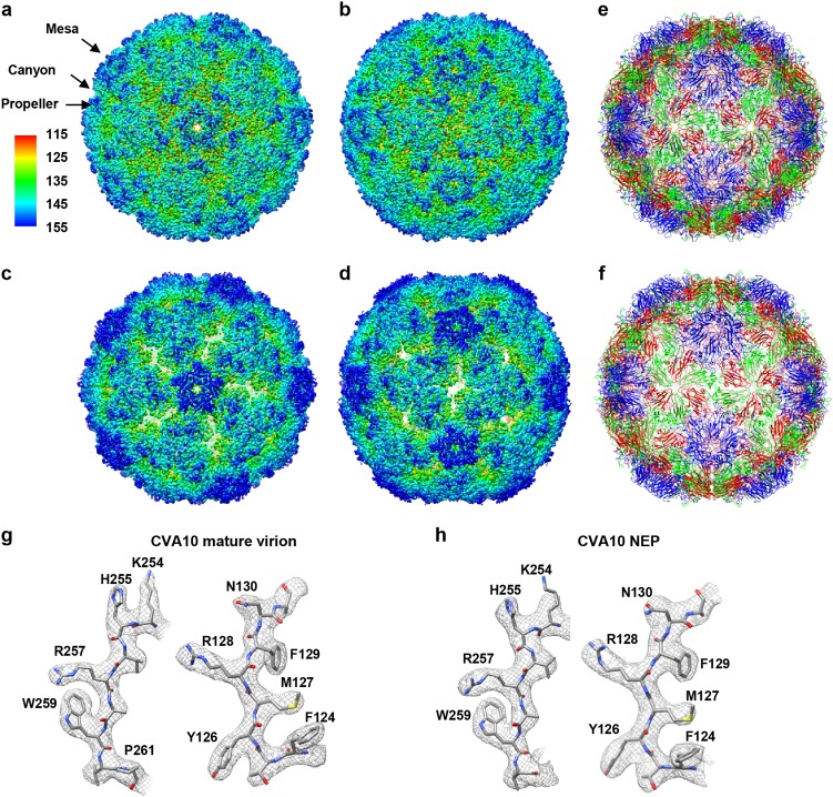
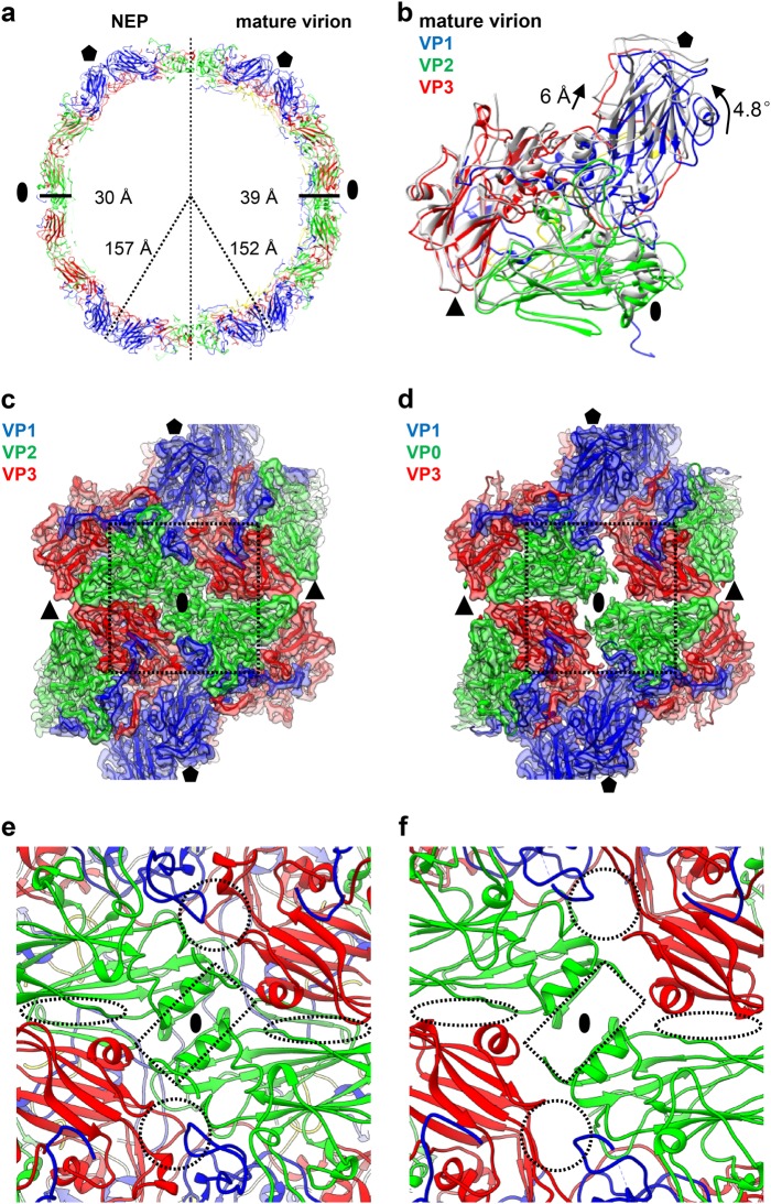
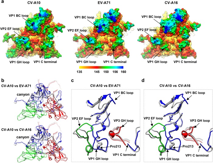
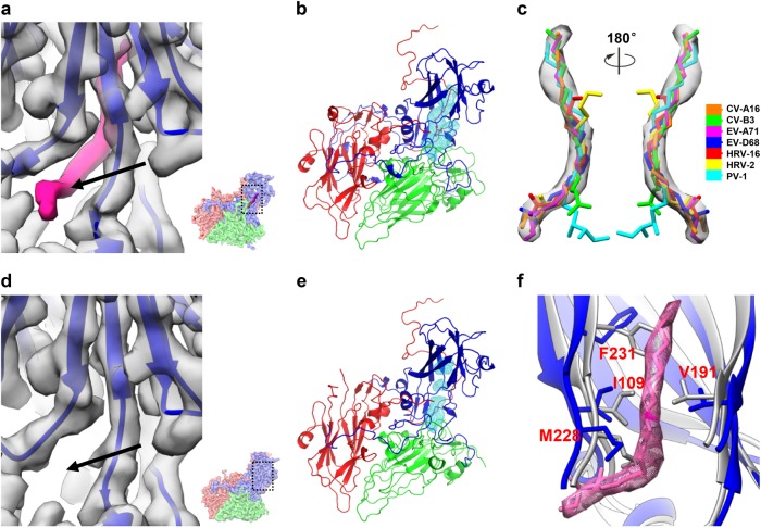
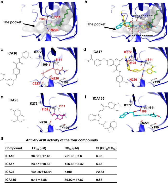
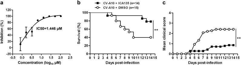
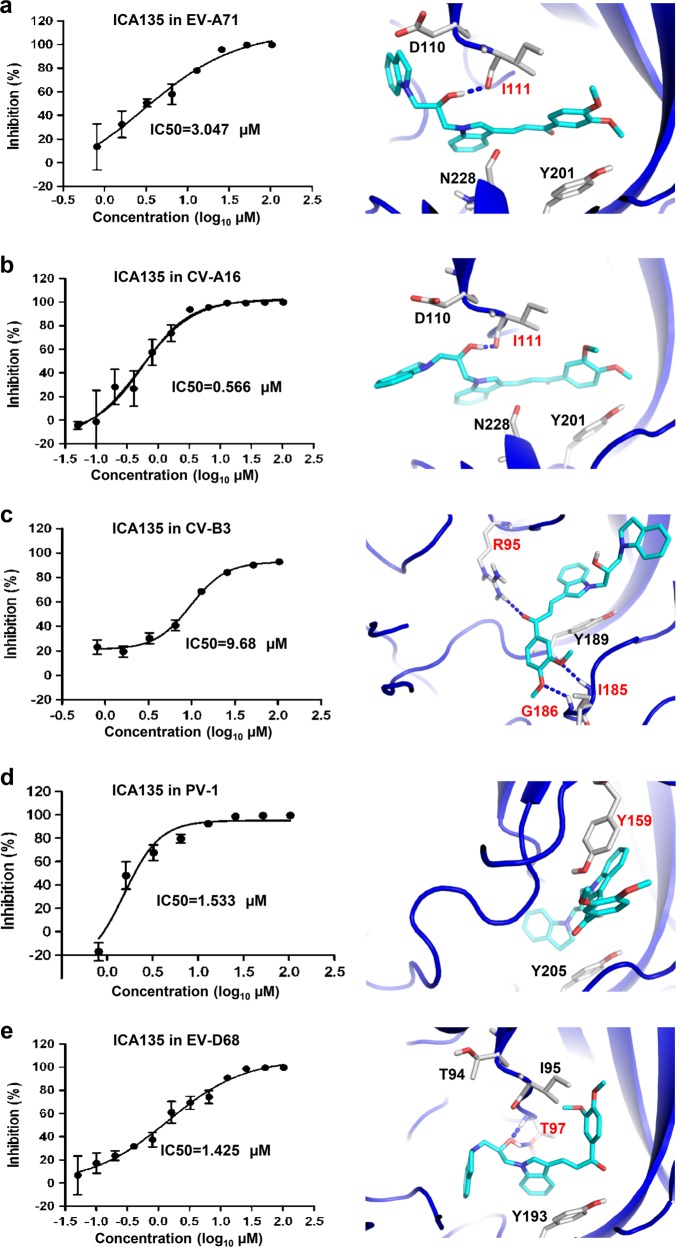
Coxsackievirus A10 (CV-A10) belongs to the Enterovirus species A and is a causative agent of hand, foot, and mouth disease. Here we present cryo-EM structures of CV-A10 mature virion and native empty particle (NEP) at 2.84 and 3.12 Å, respectively. Our CV-A10 mature virion structure reveals a density corresponding to a lipidic pocket factor of 18 carbon atoms in the hydrophobic pocket formed within viral protein 1. By structure-guided high-throughput drug screening and subsequent verification in cell-based infection-inhibition assays, we identified four compounds that inhibited CV-A10 infection in vitro. These compounds represent a new class of anti-enteroviral drug leads. Notably, one of the compounds, ICA135, also exerted broad-spectrum inhibitory effects on a number of representative viruses from all four species (A-D) of human enteroviruses. Our findings should facilitate the development of broadly effective drugs and vaccines for enterovirus infections.


Conflict of interest statement

The authors declare that they have no conflict of interest.

Figures

Fig. 1
Fig. 1. Atomic-resolution cryo-EM structures of CV-A10 particles.
a, b Cryo-EM maps of CV-A10 mature virion viewed along the icosahedral five-fold (a) and two-fold (b) axis, respectively. The color bar labels the corresponding radius from the center of the sphere (unit in Å). The same color scheme was followed throughout. c, d Cryo-EM maps of CV-A10 NEP viewed along the icosahedral five-fold (c) and two-fold (d) axis, respectively. e, f Atomic models of CV-A10 mature virion (e) and NEP (f) viewed along the icosahedral two-fold axis, respectively. The models of capsid proteins VP1, VP2, VP3, and VP4 were colored in blue, green, red, and yellow, respectively. The same color scheme was followed throughout, unless otherwise indicated. g, h Atomic-resolution structural features of CV-A10 mature virion (g) and NEP (h), respectively, with the segmented density (mesh) in gray and the corresponding atomic model (sticks) in color. The well-resolved densities for almost all the side chains demonstrate the high resolution of the cryo-EM map
Fig. 2
Fig. 2. Capsid structural comparison between CV-A10 mature virion and NEP.
a Two half sections of a 20 Å-thick central slab through the atomic models of CV-A10 NEP particle (left) and mature virion (right). Black oval and pentagon represent the two-fold and five-fold axes, respectively. The capsid radiuses and thickness for the two types of particles are also labeled. b One protomeric unit of CV-A10 NEP (in gray) was aligned with that of mature virion (in color). The rotation and translation from mature virion to NEP were also labeled. Black oval, triangle, and pentagon represent the two-fold, three-fold, and five-fold axes, respectively. c, d Structural configurations of four adjacent protomers around the two-fold axis for CV-A10 mature virion (c) and NEP (d), respectively. The major differences between them were indicated by dashed rectangle. e, f Zoom-in view of the icosahedral two-fold region of CV-A10 mature virion (e) and NEP (f). Dashed rectangle, circle, and oval indicate the locations of two-fold channel, a second channel nearby the quasi-three-fold axis, and another small ditch formed at the VP2/VP3 interface between adjacent protomers nearby the three-fold axis arising in the NEP, respectively
Fig. 3
Fig. 3. Comparison of canyon region among CV-A10, CV-A16, and EV-A71.
a Molecular surfaces of one asymmetric unit plus an adjacent VP1 within a pentamer of CV-A10 mature virion, EV-A71 (3VBF), and CV-A16 (5C4W) visualized from outside of the capsid. The surface is radially colored as indicated by the color bar. b Model alignment of one asymmetric unit and the C-terminus of an adjacent VP1 within a pentamer of CV-A10 mature virion with that of EV-A71(3VBF) (upper panel), or CV-A16 (5C4W) (lower panel). The canyon location is indicated by dashed line. c, d Zoomed-in view of the major differences in the exposed loop regions surrounding the canyon between CV-A10 and EV-A71 (c) or between CV-A10 and CV-A16 (d). The models of EV-A71 and CV-A16 are in gray, and that of CV-A10 mature virion in color. For CV-A10 mature virion, the BC loop, GH loop, and C-terminus of VP1, EF loop of VP2, and GH loop of VP3 are indicated by black arrows. Pro 213 of VP1 in CV-A10 is also indicated by a black arrow
Fig. 4
Fig. 4. VP1 pocket region and identification of pocket factor in the CV-A10 mature virion.
a Zoomed view of the VP1 pocket region in CV-A10 mature virion. The pocket location with respect to the complete CV-A10 protomer is illustrated in a small panel in the lower-right corner. The density corresponding to the pocket factor is highlighted in hot-pink. The entrance of the pocket is indicated by a black arrow. b The hydrophobic pocket (cyan mesh) in VP1 of CV-A10 mature virion is occupied by a natural lipid (hot-pink). c Fitting of putative pocket factor structures from seven known enterovirus structures (CV-A16: 5C4W, CV-B3: 1COV, EV-A71: 3VBF, EV-D68: 4WM8, HRV-16: 1AYN, HRV-2: 1FPN, and PV-1: 1VBD) into the corresponding density in the cryo-EM map of CV-A10 mature virion. d Zoom-in view of the VP1 pocket region in CV-A10 NEP. The same visualization style as in (a) was adapted. No density corresponding to the pocket factor can be observed. e The empty, collapsed pocket (cyan mesh) of CV-A10 NEP. f Comparison of the pocket region between CV-A10 mature virion (blue) and NEP (gray). Here the density and atomic model corresponding to the pocket factor are shown in hot pink
Fig. 5
Fig. 5. Binding modes of SPH and the four pocket-binding compounds and the anti-CV-A10 activity measurement.
a The pocket factor of CV-A10, SPH, occupies part of the pocket (shown as transparent surface) and forms hydrogen bonds with I109, I111, and N226 of VP1 in CV-A10. Hydrogen bonds are shown as blue dashed lines. b Predicted binding positions of the four compounds in VP1 pocket of CV-A10 mature virion. The pocket (transparent surface) and the pocket factor are also shown to illustrate the relative locations of the four compounds. The four compounds are shown as sticks and colored by elements, with the carbon atoms of ICA16, ICA17, ICA25, and ICA135 colored in salmon, yellow, magenta, and cyan, respectively. The overlapping region inside the pocket among the CV-A10 pocket factor and the four compounds is indicated by the dashed rectangle. cf Binding modes of ICA16 (c), ICA17 (d), ICA25 (e), and ICA135 (f) to the VP1 pocket of CV-A10 mature virion predicted by docking analysis (using Glide 6.9 in its SP mode). CV-A10 is shown in blue cartoon, and the compounds in stick. Potential hydrogen bonds formed between the pocket and the compounds are shown as blue dashed lines, and the residues involved in the hydrogen bond formation are labeled in red. g List of EC50, CC50, and SI (CC50/EC50) values of the four compounds against CV-A10 (S0148b). 50 µl of serially diluted compounds was mixed with 50 µl of CV-A10 virus containing 100TCID50. The mixtures were incubated for 1 h and then added to 2 × 104 RD cells, followed by incubation at 37 °C for 48 h. Then, culture supernatants were collected and analyzed for virus titers by plaque assays. Each experiment was performed at least three times
Fig. 6
Fig. 6. In vitro and in vivo inhibitory activities of ICA135 against CV-A10 infection.
a Inhibitory effect of ICA135 on CV-A10 infection of RD cells. 50 µl of CV-A10/S0148b virus containing 100 TCID50 was mixed with 50 µl of compound dilutions, and incubated at 37 °C for 1 h. The mixtures were added to 2 × 104 RD cells to allow infection at 37 °C for 6 h. Then, the mixtures were replaced with fresh medium and the plates were incubated at 37 °C for 24 h. The cells and medium were harvested and subjected to realtime PCR assay. Each experiment was performed at least three times. b, c In vivo inhibitory effect of ICA135. Groups of 7-day-old ICR mice (n = 14 or 15) were inoculated intraperitoneally (i.p.) with 8 × 104 TCID50 of CV-A10/S0148b in the absence or presence of 50 mg/kg body weight of ICA135. Then the mice were monitored daily for survival (b) and clinical signs (c) for a period of 14 days. Clinical scores were graded as follows: 0, healthy; 1, reduced mobility; 2, limb weakness; 3, paralysis; 4, death. Survival curves for the control and treatment groups were compared by Log-rank test using GraphPad Prism software. Mean clinical scores for the two groups were compared by two-way ANOVA test. Statistical significance was indicated as **P ≤ 0.01
Fig. 7
Fig. 7. Broad-spectrum anti-enterovirus activity of ICA135.
ae ICA135 was tested for its in vitro inhibitory activity against a panel of enteroviruses, including EV-A71 strain G082 (a), CV-A16 strain SZ05 (b), CV-B3 strain Nancy (c), PV1 strain Sabin (d), and EV-D68 strain US/MO/14-18947 (e). Data shown in the left panels are representative results from at least two independent experiments for each virus. Binding modes of ICA135 into the corresponding viruses (right panels) were predicted by docking analysis. EV-A71 (PDB 3VBF), CV-A16 (PDB 5C4W), EV-D68 (PDB 4WM8), CV-B3 (PDB 1COV), and PV-1 (PDB 1VBD) are shown in blue cartoon with ICA135 in stick. Potential hydrogen bonds are shown as blue dashed lines. The residues involved in the hydrogen bond formation or π–π stacking are labeled in red

Similar articles

Cited by

References

    1. Tapparel C, Siegrist F, Petty TJ, Kaiser L. Picornavirus and enterovirus diversity with associated human diseases. Infect. Genet. Evol. 2013;14:282–293. doi: 10.1016/j.meegid.2012.10.016. - DOI - PubMed
    1. Yamashita T, Ito M, Taniguchi A, Sakae K. Prevalence of coxsackievirus A5, A6, and A10 in patients with herpangina in Aichi Prefecture, 2005. Jpn. J. Infect. Dis. 2005;58:390–391. - PubMed
    1. Mirand A, et al. Outbreak of hand, foot and mouth disease/herpangina associated with coxsackievirus A6 and A10 infections in 2010, France: a large citywide, prospective observational study. Clin. Microbiol. Infect. 2012;18:E110–E118. doi: 10.1111/j.1469-0691.2012.03789.x. - DOI - PubMed
    1. Lu QB, et al. Circulation of Coxsackievirus A10 and A6 in hand-foot-mouth disease in China, 2009–2011. PLoS One. 2012;7:e52073. doi: 10.1371/journal.pone.0052073. - DOI - PMC - PubMed
    1. He YQ, et al. Emergence, circulation, and spatiotemporal phylogenetic analysis of coxsackievirus A6-and coxsackievirus A10-associated hand, foot, and mouth disease infections from 2008 to 2012 in Shenzhen, China. J. Clin. Microbiol. 2013;51:3560–3566. doi: 10.1128/JCM.01231-13. - DOI - PMC - PubMed