. 2018 Nov 28;9(1):53-73.
doi: 10.1002/2211-5463.12542. eCollection 2019 Jan.

Glyceraldehyde-3-phosphate dehydrogenase from Citrobacter sp. S-77 is post-translationally modified by CoA (protein CoAlation) under oxidative stress

Affiliations

Glyceraldehyde-3-phosphate dehydrogenase from Citrobacter sp. S-77 is post-translationally modified by CoA (protein CoAlation) under oxidative stress

Kohsei Tsuji et al. FEBS Open Bio. .

Abstract

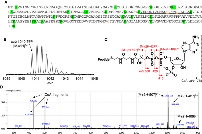
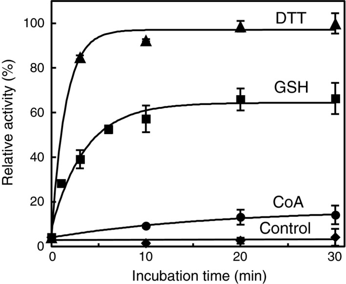
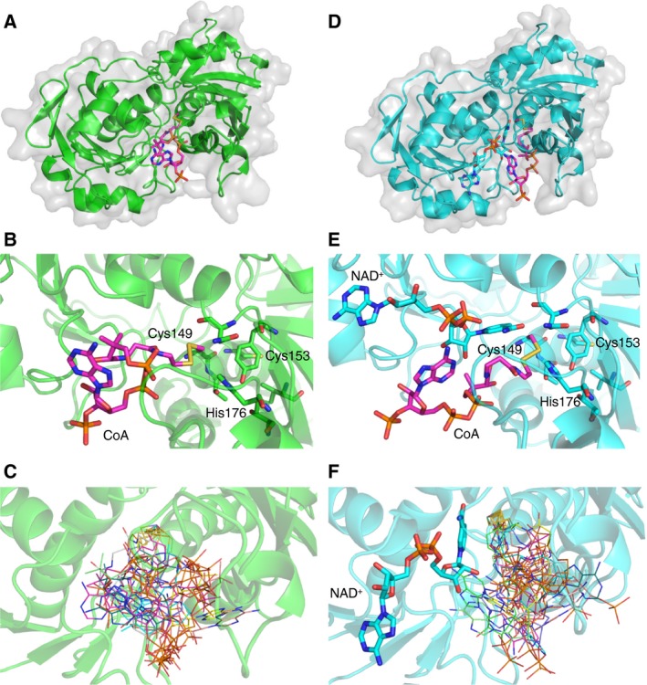
Protein CoAlation (S-thiolation by coenzyme A) has recently emerged as an alternative redox-regulated post-translational modification by which protein thiols are covalently modified with coenzyme A (CoA). However, little is known about the role and mechanism of this post-translational modification. In the present study, we investigated CoAlation of glyceraldehyde-3-phosphate dehydrogenase (GAPDH) from a facultative anaerobic Gram-negative bacterium Citrobacter sp. S-77 (Cb GAPDH). GAPDH is a key glycolytic enzyme whose activity relies on the thiol-based redox-regulated post-translational modifications of active-site cysteine. LC-MS/MS analysis revealed that CoAlation of Cb GAPDH occurred in vivo under sodium hypochlorite (NaOCl) stress. The purified Cb GAPDH was highly sensitive to overoxidation by H2O2 and NaOCl, which resulted in irreversible enzyme inactivation. By contrast, treatment with coenzyme A disulphide (CoASSCoA) or H2O2/NaOCl in the presence of CoA led to CoAlation and inactivation of the enzyme; activity could be recovered after incubation with dithiothreitol, glutathione and CoA. CoAlation of the enzyme in vitro was confirmed by mass spectrometry. The presence of a substrate, glyceraldehyde-3-phosphate, fully protected Cb GAPDH from inactivation by CoAlation, suggesting that the inactivation is due to the formation of mixed disulphides between CoA and the active-site cysteine Cys149. A molecular docking study also supported the formation of mixed disulphide without steric constraints. These observations suggest that CoAlation is an alternative mechanism to protect the redox-sensitive thiol (Cys149) of Cb GAPDH against irreversible oxidation, thereby regulating enzyme activity under oxidative stress.

Keywords: S‐thiolation; coenzyme A; glyceraldehyde‐3‐phosphate dehydrogenase; post‐translational modification; redox regulation.


Figures

Figure 1
Figure 1
In vivo CoAlation of Cb GAPDH under NaOCl stress. (A) The predicted cleavage sites in the sequence of Cb GAPDH after Lys‐C digestion are depicted in green background, and the resulting peptides containing Cys149, Cys153 and Cys288 (Y138–K159 and G269–K295) are underlined with Cys residues coloured by orange character. (B) The precursor ion of CoAlated peptide (m/z 1040.763+) (C) Schematic illustration of fragmentations of CoA by CID during MS/MS spectra acquisitions. Neutral loss of precursor ions characteristic to the loss of CoA fragments (m/z 410, 428, 508) by CID served for verification of the CoAlated peptides. (D) The MS/MS spectrum of in vivo CoAlated peptide of Cb GAPDH under NaOCl stress. The abundance of the peaks of precursor ions minus CoA fragments led to decreased intensity of y‐ and b‐ion series, which hampered identification of the peptide backbone fragmentations. The results presented here are one of the representatives of two independent experiments.
Figure 2
Figure 2
Inactivation profile of Cb GAPDH by CoASSCoA and GSSG. (A) Time‐course inactivation of Cb GAPDH by CoASSCoA. The enzyme was incubated with 1 mm CoASSCoA (■), 1 mm CoA (○) or buffer as control (◆) for 0–15 min. (B) Time‐dependent inactivation of Cb GAPDH by CoASSCoA, 250 μm (●), 500 μm (▲), 750 μm (◆) and 1000 μm (■). (Inset) Concentration dependence of the pseudo‐first‐order rate constant of enzyme inactivation. (C) Time‐dependent inactivation of Cb GAPDH by GSSG, 2.5 mm (●), 5.0 mm (▲), 7.5 mm (◆) and 10 mm (■). (Inset) Concentration dependence of the pseudo‐first‐order rate constant of enzyme inactivation. Activities are given as a percentage of the initial activity (100 ± 5.3 U·mg−1) before the inactivation experiment. The results are presented as means of at least three independent experiments with standard deviations.
Figure 3
Figure 3
Reversibility of Cb GAPDH by DTT and effects of the substrate and cofactor on inactivation of Cb GAPDH. (A) Reversibility of the inactivated enzyme by DTT. The Cb GAPDH inactivated by 1 mm CoASSCoA for 15 min was incubated with 10 mm DTT for 15 min, and then, residual activity was assessed before (black bar) and after (white bar) treatment of DTT. The control experiment (without any treatment) is given for comparison. (B) The effects of G3P or NAD + during inactivation treatment. The enzyme was preincubated with 2 mm G3P (♢) or 1 mm NAD + (▵) for 5 min and then treated with 1 mm CoASSCoA for 0–15 min. Inactivation curve of 1 mm CoASSCoA is presented for comparison (■). Activities are given as a percentage of the initial activity (100 ± 5.3 U·mg−1) before the inactivation experiment. The results are presented as means of at least three independent experiments with standard deviations.
Figure 4
Figure 4
MALDITOF mass spectra of Cb GAPDH treated with CoASSCoA. (A) The spectrum of native enzyme was acquired without any treatment. The spectra of Cb GAPDH incubated with 1 mm CoASSCoA (30 min) were acquired before (B) and after (C) the treatment with 10 mm DTT (30 min). The signals marked by * are attributed to the adducts of sinapinic acid.
Figure 5
Figure 5
Identification of modification sites of in vitro CoAlated Cb GAPDH by using LCMS/MS. (A) The fragmentation pattern of MS/MS spectrum indicates that active‐site Cys149 is CoAlated, while Cys153 is carbamidomethylated. (B) Intramolecular disulphide bond between Cys149 and 153 was also detected as an additional redox modification of Cb GAPDH. (C) These redox modifications were reversed in a DTT sensitive manner, which suggested the reversibility of these redox post‐translational modifications.
Figure 6
Figure 6
Inactivation profile of Cb GAPDH by H2O2 with/without CoA and the reversibility by DTT. (A) Time‐course inactivation of Cb GAPDH by H2O2 with/without G3P or NAD +. The enzyme was incubated with only 0.1 mm H2O2 (●), plus 2 mm G3P (◆) or plus 1 mm NAD + (▲) for 0–15 min. G3P or NAD + was preincubated with enzyme for 5 min before the treatment with H2O2. (B) Time‐course inactivation of Cb GAPDH by H2O2 plus CoA with/without G3P or NAD +. The enzyme was incubated with only 0.1 mm H2O2 and 1 mm CoA (○), plus 2 mm G3P (♢) or plus 1 mm NAD + (▵) for 0–15 min. CoA, G3P and/or NAD + were preincubated with the enzyme for 5 min before the treatment with H2O2. (C) Reversibility of the inactivated enzyme by DTT. The Cb GAPDH inactivated at indicated conditions for 15 min was incubated with 10 mm DTT for 15 min, and then, residual activity was assessed before (black bar) and after (white bar) treatment with DTT. The control experiment (without any treatment) is given for comparison. Activities are given as a percentage of the initial activity (100 ± 5.3 U·mg−1) before the inactivation experiment. The results are presented as means of at least three independent experiments with standard deviations.
Figure 7
Figure 7
MALDITOF mass spectra of Cb GAPDH treated with H2O2 plus CoA. The spectra of the enzyme incubated with 0.1 mm H2O2 plus 1 mm CoA (30 min) were acquired before (A) and after (B) the treatment with 10 mm DTT (30 min). The signals marked by * are assigned to the adducts of sinapinic acid.
Figure 8
Figure 8
Time‐course reactivation of CoAlated Cb GAPDH by thiol compounds. The CoAlated enzyme was incubated with 10 mm DTT (▲), 5 mm GSH (■), 1 mm CoA (●) and buffer alone (◆) for 0–30 min, and the remaining activity was assessed. Activities are given as a percentage of the maximum activity measured after 30 min incubation with 10 mm DTT (93.5 ± 5.0 U·mg−1). The results are presented as means of at least three independent experiments with standard deviations.
Figure 9
Figure 9
Quantification of free thiols and pKa determination. (A) The number of free thiols in native, oxidised and CoAlated Cb GAPDH. The absorbance at 412 nm of the solution after the reaction with free thiols in Cb GAPDH and DTNB was measured by UV–vis. (B) pKa titration curve of the active‐site Cys149 in Cb GAPDH. The pKa was estimated to be 5.82 ± 0.05 by fitting each point to a derivation of the Henderson–Hasselbalch equation. The results are presented as means of at least three independent experiments with standard deviations.
Figure 10
Figure 10
Homology‐modelled structure of Cb GAPDH. (A) Overall and (B) active‐site structure of apo Cb GAPDH. Cys149, Cys153, Cys288 and His176 are depicted by orange and violet sticks. The broken lines represent the distances between two sulfur or nitrogen atoms.
Figure 11
Figure 11
Molecular docking of Cb GAPDH and CoA by using the flexible side chain method in autodock4. The best poses of the modelled covalent complex between CoA and Cys149 of apo (A–C) or holo (D–F) Cb GAPDH are shown. The estimated free energies of the best binding pose were −10.37 and −8.38 kcal·mol−1 for the apo and holo structures, respectively. CoA is depicted in pink in the overall docking pose (A, D) and active site (B, E). The 10 best poses were overlaid and depicted in lines with different colours (C, F).
Figure 12
Figure 12
Schematic illustration of the CoAlation and de‐CoAlation mechanisms of Cb GAPDH. The active‐site Cys149 of Cb GAPDH forms cysteine sulphenate or sulphenyl chloride that further undergo overoxidation to sulfonate or intramolecular disulphide bonding in the presence of H2O2 or NaOCl alone, which resulted in irreversible inactivation of the glycolytic activity. While, Cb GAPDH is protected against this irreversible overoxidation by protein CoAlation and intramolecular disulphide bonding in the presence of H2O2/NaOCl and CoA. These modifications resulted in reversible enzyme inactivation due to CoAlation of Cys149.

Similar articles

Cited by

References

    1. Loi VV, Rossius M and Antelmann H (2015) Redox regulation by reversible protein S‐thiolation in bacteria. Front Microbiol 6, 187. - PMC - PubMed
    1. Grek CL, Zhang J, Manevich Y, Townsend DM and Tew KD (2013) Causes and consequences of cysteine S‐glutathionylation. J Biol Chem 288, 26497–26504. - PMC - PubMed
    1. Antelmann H and Hamilton CJ (2012) Bacterial mechanisms of reversible protein S‐thiolation: structural and mechanistic insights into mycoredoxins. Mol Microbiol 86, 759–764. - PubMed
    1. Brandes N, Schmitt S and Jakob U (2009) Thiol‐based redox switches in eukaryotic proteins. Antioxid Redox Signal 11, 997–1014. - PMC - PubMed
    1. Van Laer K, Hamilton CJ and Messens J (2013) Low‐molecular‐weight thiols in thiol‐disulfide exchange. Antioxid Redox Signal 18, 1642–1653. - PubMed

Publication types

MeSH terms

Substances