

Imbalance in gut microbes from babies born to obese mothers increases gut permeability and myeloid cell adaptations that provoke obesity and NAFLD
- PMID: 30652107
- PMCID: PMC6334233
- DOI: 10.15698/mic2019.01.666
Imbalance in gut microbes from babies born to obese mothers increases gut permeability and myeloid cell adaptations that provoke obesity and NAFLD
Abstract
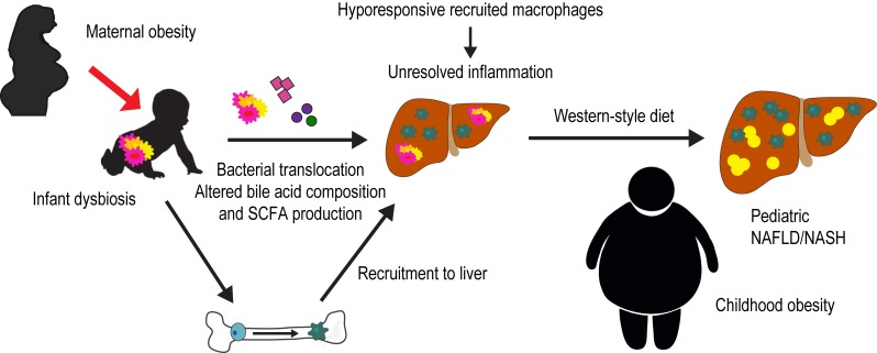
Non-alcoholic fatty liver disease (NAFLD) is a multifactorial disease affecting nearly 40% of obese youth and up to 10% of the general pediatric population. A key aspect of NAFLD pathogenesis is proinflammatory hepatic macrophage activation and hepatic recruitment of circulating monocytes, which originate from the bone marrow. In neonates, the activation and polarization of myeloid immune cells are normally shaped in part by systemic factors derived from intestinal microbiota during the first 1000 days of life. Perturbations of the gut microbiome, and in turn the metabolites and bacterial products released systemically, can affect the functional phenotype of these immune cells. Evidence in germ-free mice has shown that fecal microbial transfer from obese mice or obese human donors promotes obesity and inflammation in the recipients, suggesting a direct role for the gut microbiome in promoting obesity and possibly NAFLD. Indeed, patients suffering from NAFLD show evidence for dysbiosis, increased gut permeability, and changes in bile acids that drive the progression of hepatic inflammation toward non-alcoholic steatohepatitis (NASH), the more severe form of the disease. Compared with infants born to normal-weight mothers, we previously showed that the gut microbiome from neonates born to obese mothers is compositionally distinct. However, whether this alteration in early gut microbiota in infants born to obese mothers can cause inflammatory processes that initiate development of NAFLD or obesity is unknown. How these alterations contribute to long-term immune cell mediated liver inflammation and progression of NAFLD needs to be determined. Our recently published work (Soderborg et al., Nat Commun 9:4462) demonstrates a causative role of early life microbiome dysbiosis in infants born to mothers with obesity in novel pathways that promote developmental programming of NAFLD.
Conflict of interest statement
Conflict of interest: J.E.F. is a consultant to the scientific advisory board of Janssen Pharmaceuticals.
Figures
Comment on
-
The gut microbiota in infants of obese mothers increases inflammation and susceptibility to NAFLD.Nat Commun. 2018 Oct 26;9(1):4462. doi: 10.1038/s41467-018-06929-0. Nat Commun. 2018. PMID: 30367045 Free PMC article.
Publication types
Grants and funding
LinkOut - more resources
Full Text Sources
Other Literature Sources