. 2019 Jan 8:2:15.
doi: 10.1038/s42003-018-0264-7. eCollection 2019.

Optogenetic control of integrin-matrix interaction

Affiliations

Optogenetic control of integrin-matrix interaction

Julia Baaske et al. Commun Biol. .

Abstract

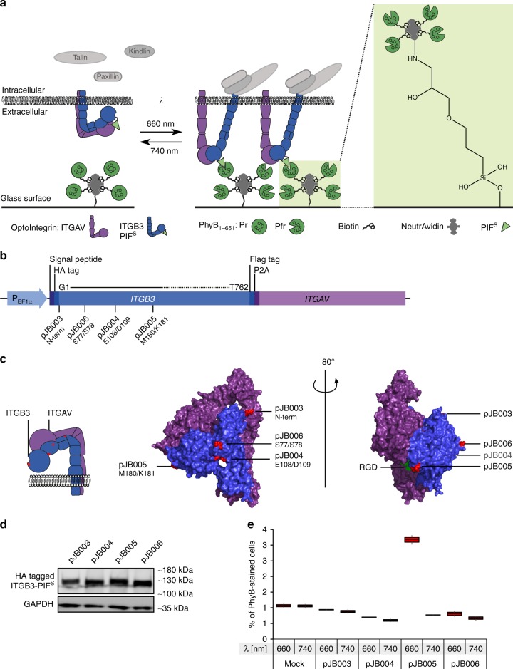
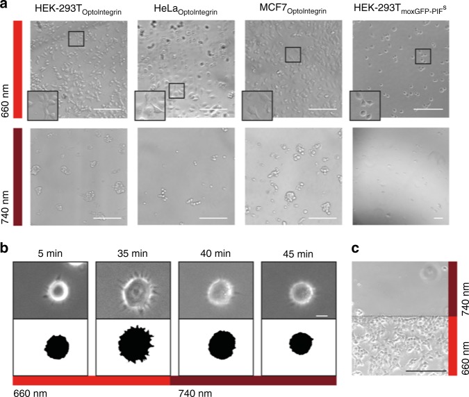
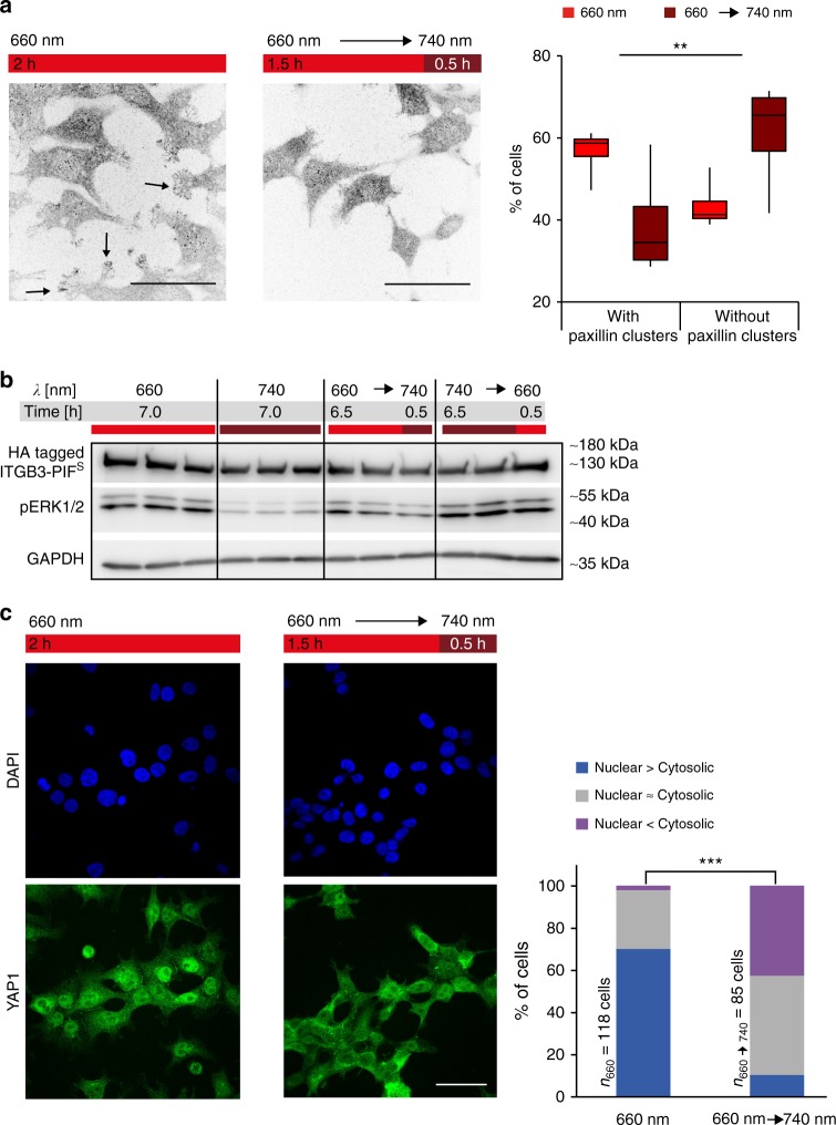
Optogenetic approaches have gathered momentum in precisely modulating and interrogating cellular signalling and gene expression. The use of optogenetics on the outer cell surface to interrogate how cells receive stimuli from their environment, however, has so far not reached its full potential. Here we demonstrate the development of an optogenetically regulated membrane receptor-ligand pair exemplified by the optically responsive interaction of an integrin receptor with the extracellular matrix. The system is based on an integrin engineered with a phytochrome-interacting factor domain (OptoIntegrin) and a red light-switchable phytochrome B-functionalized matrix (OptoMatrix). This optogenetic receptor-ligand pair enables light-inducible and -reversible cell-matrix interaction, as well as the controlled activation of downstream mechanosensory signalling pathways. Pioneering the application of optogenetic switches in the extracellular environment of cells, this OptoMatrix-OptoIntegrin system may serve as a blueprint for rendering matrix-receptor interactions amendable to precise control with light.


Conflict of interest statement

The authors declare no competing interests.

Figures

Fig. 1
Fig. 1
Design and construction of light-inducible receptor–ligand interactions. a (3-glycidyloxypropyl)trimethoxysilane (GLYMO)-functionalized glass slides are coated with NeutrAvidin and subsequently with biotinylated PhyB1–651. Cells expressing PIFS-coupled OptoIntegrins are seeded on top of glass slides. Upon illumination with 660 nm light, PhyB1–651 converts from its inactive Pr form to the active Pfr form. The active PhyB1–651 form interacts with the OptoIntegrin which then activates mechanosensory pathways. b Schematic view of the OptoIntegrin-encoding sequence with PIFS insertion sites. Amino acids numbers under plasmid names refer to insertion sites in the crystal structure (Protein data bank accession number: 1L5G). c Crystal structure of αVβ3 extracellular domain with RGD ligand (1L5G). ITGB3 subunit in blue and ITGAV subunit in purple. PIFS insertion sites for different constructs are marked in red. d Expression of different OptoIntegrin constructs in HEK-293T cells. Western blot analysis for presence of HA-tagged β subunit in cell lysate expressing different integrin constructs. e PhyB1–651 binding to surface of untransfected (Mock) HEK-293T cells and HEK-293T cells transiently expressing OptoIntegrin constructs under illumination with 660 nm or 740 nm light. Analysis of stained cells with flow cytometry. Data of two replicates (n > 5000 cells) per condition is presented in box and whisker plot format
Fig. 2
Fig. 2
Validation of cell–matrix interaction with different cell lines stably expressing OptoIntegrin. a HEK-293T, HeLa and MCF7 cells stably expressing OptoIntegrin or moxGFP-PIFS were seeded on PhyB1–651-coated glass slides and incubated under 660 nm or 740 nm light (I = 20 µmol m−2 s−1) for 5 h and subsequently imaged using a transmission light microscope (scale bar = 200 µm). b Live cell imaging of cell–matrix interaction under 660 nm light and then switch to 740 nm light after 35 min with binary images of cell shapes to illustrate the cells spreading. For this, HeLa cells stably expressing OptoIntegrin were seeded on OptoMatrix and subsequently imaged. Micrographs were taken at indicated time points (scale bar = 10 µm). c Spatial control of cell attachment with OptoIntegrin-expressing HEK-293T cells. OptoIntegrin-expressing cells were cultivated on OptoMatrix, locally illuminated with 660 nm or 740 nm for 3 min and then left in darkness for 4 h. Afterwards, cells were fixed and imaged (scale bar = 200 µm)
Fig. 3
Fig. 3
Transducing signals from the extracellular to the intracellular with cells expressing OptoIntegrin. Cells were seeded on PhyB1–651-coated glass surface and illuminated as indicated. a Paxillin staining with anti-paxillin antibody of fixed stably OptoIntegrin-expressing HEK-293T cells. The intensities of the images were inverted to better visualize clusters at cell edges. The data of three independent experiments where at least 35 cells per condition were analysed in a blinded manner is presented in box and whisker plot format (p = 0.000989 using Cochran–Mantel–Haenszel-test with one degree of freedom, showing a consistent difference in the populations across several repeats). b Phosphorylation of ERK1/2 in stably OptoIntegrin-expressing HEK-293T cells in response to different light conditions analysed by western blot. Biological triplicates are shown for each condition using anti-pERK1/2 antibodies and anti-HA antibodies to detect pERK1/2 and OptoIntegrin HA tagged β subunit, respectively. c YAP1 (green) and nucleus (DAPI, blue) staining of OptoIntegrin-expressing HEK-293T cells. Chart shows the percentage of cells from a single experiment where YAP1 is localized mainly in the cytoplasm, mainly in the nucleus or in an equal distributions between cytoplasm and nucleus (p < 0.0001, with a Chi-square value of 87.56 and two degrees of freedom, using Chi-square test of independence shows dependent variables). For (a), (b) and (c) λ1/2 = 660/740 nm, I1/2 = 20 µmol m−2 s−1 and for a and b scale bar = 50 µm

References

    1. Kolar K, Weber W. Synthetic biological approaches to optogenetically control cell signaling. Curr. Opin. Biotechnol. 2017;47:112–119. doi: 10.1016/j.copbio.2017.06.010. - DOI - PubMed
    1. Leopold, A. V., Chernov, K. G. & Verkhusha, V. V. Optogenetically controlled protein kinases for regulation of cellular signaling. Chem. Soc. Rev. (2018). 10.1039/C7CS00404D. - PMC - PubMed
    1. Nihongaki Y, Otabe T, Sato M. Emerging approaches for spatiotemporal control of targeted genome with inducible CRISPR-Cas9. Anal. Chem. 2018;90:429–439. doi: 10.1021/acs.analchem.7b04757. - DOI - PubMed
    1. Kolar K, Knobloch C, Stork H, Žnidarič M, Weber W. OptoBase: A web platform for molecular optogenetics. ACS Synth. Biol. 2018;7:1825–1828. doi: 10.1021/acssynbio.8b00120. - DOI - PubMed
    1. Kadem LF, et al. High-frequency mechanostimulation of cell adhesion. Angew. Chem. - Int. Ed. 2017;56:225–229. doi: 10.1002/anie.201609483. - DOI - PMC - PubMed

Publication types