. 2019 Apr 12;5(4):598-617.
doi: 10.1021/acsinfecdis.8b00345. Epub 2019 Feb 4.

Investigation of ( S)-(-)-Acidomycin: A Selective Antimycobacterial Natural Product That Inhibits Biotin Synthase

Affiliations

Investigation of ( S)-(-)-Acidomycin: A Selective Antimycobacterial Natural Product That Inhibits Biotin Synthase

Matthew R Bockman et al. ACS Infect Dis. .

Abstract

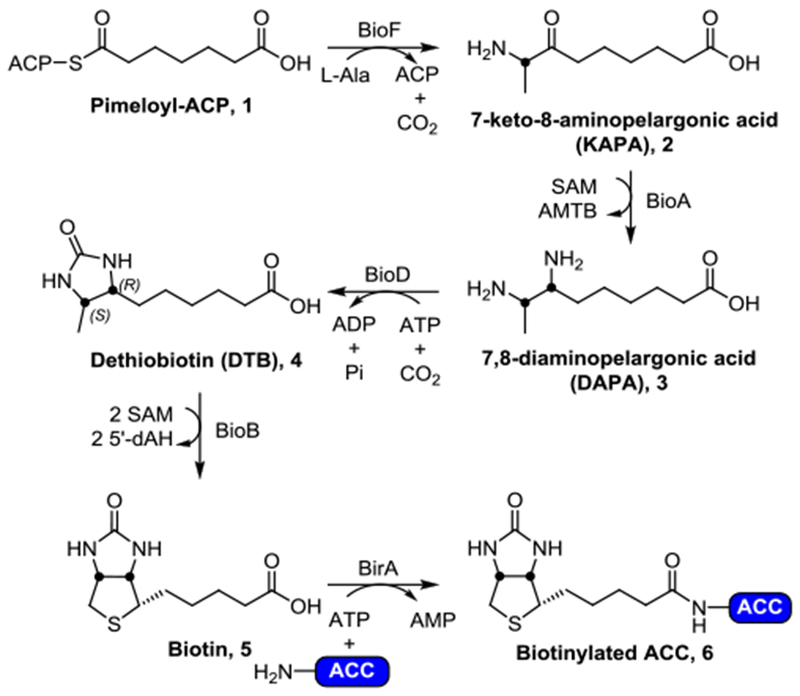
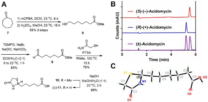
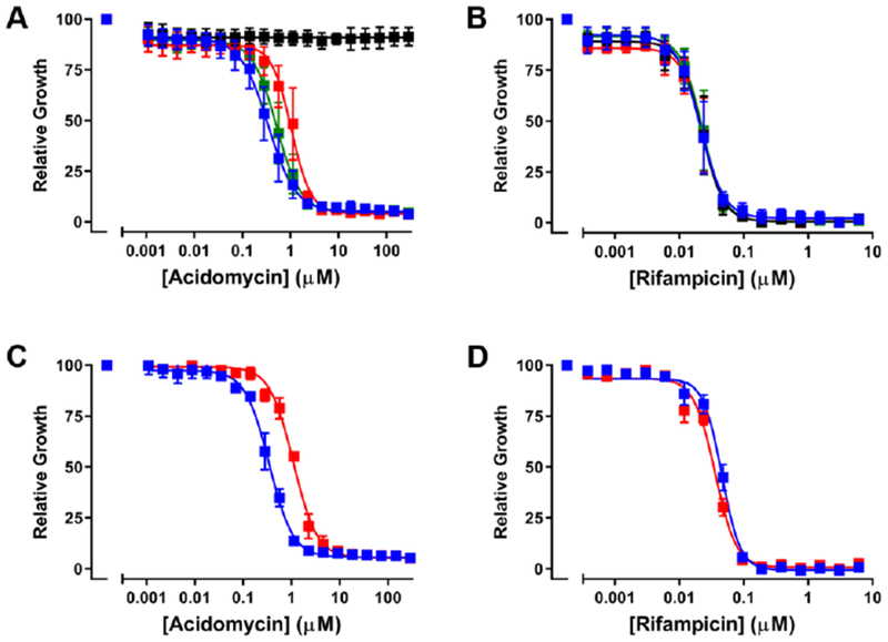
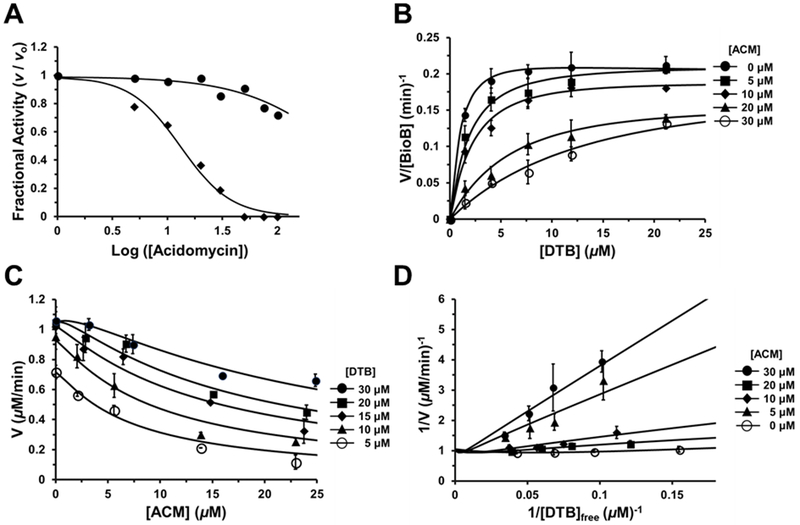
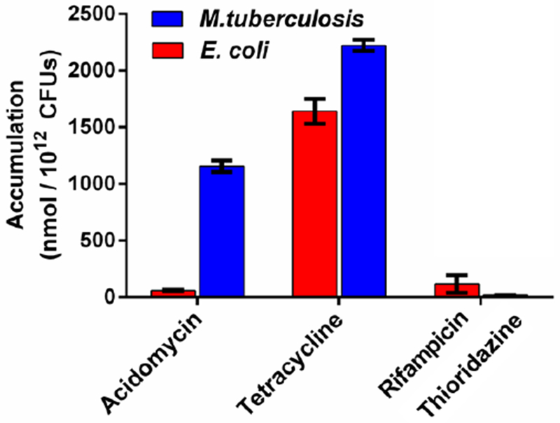
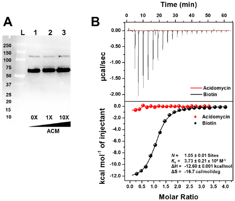
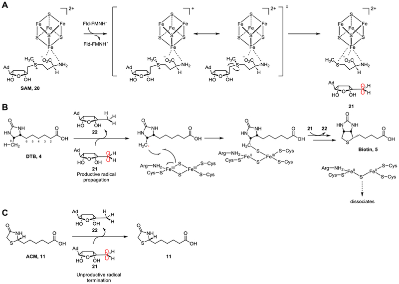
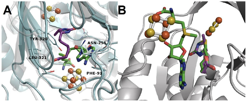
The synthesis, absolute stereochemical configuration, complete biological characterization, mechanism of action and resistance, and pharmacokinetic properties of ( S)-(-)-acidomycin are described. Acidomycin possesses promising antitubercular activity against a series of contemporary drug susceptible and drug-resistant M. tuberculosis strains (minimum inhibitory concentrations (MICs) = 0.096-6.2 μM) but is inactive against nontuberculosis mycobacteria and Gram-positive and Gram-negative pathogens (MICs > 1000 μM). Complementation studies with biotin biosynthetic pathway intermediates and subsequent biochemical studies confirmed acidomycin inhibits biotin synthesis with a Ki of approximately 1 μM through the competitive inhibition of biotin synthase (BioB) and also stimulates unproductive cleavage of S-adenosyl-l-methionine (SAM) to generate the toxic metabolite 5'-deoxyadenosine. Cell studies demonstrate acidomycin selectively accumulates in M. tuberculosis providing a mechanistic basis for the observed antibacterial activity. The development of spontaneous resistance by M. tuberculosis to acidomycin was difficult, and only low-level resistance to acidomycin was observed by overexpression of BioB. Collectively, the results provide a foundation to advance acidomycin and highlight BioB as a promising target.

Keywords: Mycobacterium tuberculosis; accumulation; acidomycin; antimetabolite; biotin biosynthesis; biotin synthase; tuberculosis.


Conflict of interest statement

The authors declare no competing financial interests.

Figures

Figure 1.
Figure 1.
The conserved biotin biosynthetic pathway. Briefly, pimeloyl-ACP (1) is converted to 7-keto-8-aminopelargonic acid (KAPA, 2) by BioF (KAPA synthetase). Transamination by BioA (DAPA synthetase) converts 2 to 7,8-diaminopelargonic acid (DAPA, 3), followed by insertion of a carbonyl by BioD (dethiobiotin synthetase) gives rise to dethiobiotin (DTB, 4). Finally, BioB (biotin synthase) is responsible for the conversion of 4 to biotin (5). The biological fate of biotin (5) is being ligated onto biotin-dependent proteins (such as acyl-CoA carboxylases, ACCs) by BirA (biotin protein ligase, BPL) affording the catalytically active biotinylated holo-ACC (6).
Figure 2.
Figure 2.
A) Synthetic route to (±)-11 (acidomycin); B) Analytical chiral HPLC trace of (±)-acidomycin (purple), with each resolved enantiomer (S)-(−)-acidomycin (red) and (R)-(+)-acidomycin (blue) using Phenomenex® Cellulose-1 5 μm 250 × 2.0 mm LC column; C) X-ray crystal structure of (−)-acidomycin. See Supporting Information for further details.
Figure 3.
Figure 3.
Impact of extrabacterial biotin and overexpression of BioB on susceptibility of M. tuberculosis H37Rv to (±)-acidomycin. Panels A and B show MIC curves for (±)-acidomycin (A) and rifampicin (B) in GAST medium supplemented with 1 μM biotin pathway intermediates (black, biotin; red, DTB; green, KAPA) or DMSO only (blue). Panels C and D show MIC curves for M. tuberculosis H37Rv (blue squares) and the BioB merodiploid strain (red squares) against (±)-acidomycin (C) and rifampicin (D) grown in GAST medium. Normalized growth was calculated at OD580 at the indicated concentration divided by the OD580 with drug (DMSO). Data are averages (± SD) of triplicate cultures and are representative of two independent experiments.
Figure 4.
Figure 4.
Inhibition of EcBioB by acidomycin. A) Concentration-response plots of the fractional initial velocity of EcBioB as a function of acidomycin concentration. (R)-(+)-acidomycin (solid circles) shows weak inhibition (IC50 of >100 μM) while the calculated IC50 for (S)-(−)-acidomycin (solid diamonds) is 13.3 ± 1.8 μM. B) Saturation curve of initial velocity of EcBioB as a function of DTB concentration at different fixed concentrations of (±)-acidomycin. Each curve was fit to Segel’s model for enzymes with multiple catalytic sites. C) Data from panel B replotted and fit to a modified version of the Morrison equation to obtain Ki for (±)-acidomycin (Ki = 2.0 ± 1.3 μM). D) Lineweaver-Burke double reciprocal plot of initial velocity versus free DTB concentration at different concentrations of (±)-acidomycin. The intersection near the y-axis exhibits some curvature because the free ligand and steady-state assumptions are not valid with the implemented assay conditions.
Figure 5.
Figure 5.
Differential accumulation in M. tuberculosis (blue bars) vs. E. coli (red bars) grown in the absence of biotin. Accumulation levels (represented in nmol / 1012 CFUs) are shown for acidomycin, high accumulating antibiotic (tetracycline) and low accumulating antibiotics (rifampicin and thioridazine).
Figure 6.
Figure 6.
A) Far-western blot of the biotinylated proteome of M. tuberculosis grown in biotin-free 7H9 media, in the presence of acidomycin. Concentration of acidomycin (μM) is depicted at the bottom of the blot. Lane L: Protein ladder; Lanes 1–3: M. tuberculosis grown in the presence of acidomycin at 0× (lane 1), 1× (lane 2) and 10× (lane 3) its MIC. This experiment was performed twice, independently. B) Isothermal titration calorimetry thermogram of (S)-(−)-acidomycin (750 μM, red) and biotin (750 μM, black) titrated into MtBPL (50 μM), with the binding data for biotin shown in black. This experiment was repeated twice, independently.
Figure 7.
Figure 7.
Mean plasma concentration versus time curves after single p.o. (25 mg/kg, blue) and i.v. (5 mg/kg, red) administration of acidomycin to CD-1 mice. Error bars represent standard deviation of the mean (n = 3); A) (R)-(+)-acidomycin; B) (S)-(−)-acidomycin.
Figure 8.
Figure 8.
Proposed mechanism of BioB and inhibition by acidomycin (11). A) A single electron from flavodoxin reduces the [4Fe–4S]+ cluster, promoting the homolytic cleavage of SAM (20) into methionine and a high-energy 5′-deoxyadenosyl radical (21). B) Under normal circumstances, the 5′-deoxyadenosyl radical (21) abstracts the C-9 hydrogen atom of the native substrate DTB (4), forming 5′-deoxyadenosine (22) and allows for the insertion of sulfur from the [2Fe–2S]+ cluster (labeled as “productive radical propagation”). Another 5′-deoxyadenosyl radical (21) is responsible for the C-6 hydrogen atom abstraction, completing the thiophane ring in biotin (5). C) When acidomycin (11) is bound to the active site instead, the lack of a C-6 hydrogen atom prevents the highly reactive 5′-deoxyadenosyl radical (21) from H-atom abstraction, leading to the radical to be unproductively quenched as a result (labeled as “unproductive radical termination”).
Figure 9.
Figure 9.
A) Homology model of EcBioB (grey, faded) aligned to MtBioB (blue, solid) depicting DTB (purple) and SAM (green) bound to the active site. All key interactions with residues remain the same between species. B) EcBioB with comparison of acidomycin and DTB (green and purple, respectively) aligned in the active site.

References

    1. WHO Global tuberculosis report; World Health Organization; Geneva, Switzerland, 2018.
    1. Brites D; Gagneux S, Co-evolution of Mycobacterium tuberculosis and Homo sapiens. Immunol. Rev. 2015, 264, 6–24. DOI: 10.1111/imr.12264. - DOI - PMC - PubMed
    1. Gagneux S, Ecology and evolution of Mycobacterium tuberculosis. Nat. Rev. Microbiol 2018, 16, 202–213. DOI: 10.1038/nrmicro.2018.8. - DOI - PubMed
    1. Organization W. H., Treatment of tuberculosis. Guidelines. 4th ed.; WHO/HTM/TB/2009.420: 2010. - PubMed
    1. Wohlleben W; Mast Y; Stegmann E; Ziemert N, Antibiotic drug discovery. Microb. Biotechnol 2016, 9, 541–548. DOI: 10.1111/1751-7915.12388. - DOI - PMC - PubMed

Publication types

MeSH terms

LinkOut - more resources