. 2019 Nov 1;31(13):917-932.
doi: 10.1089/ars.2018.7664. Epub 2019 Mar 20.

Gender Difference in Damage-Mediated Signaling Contributes to Pulmonary Arterial Hypertension

Affiliations

Gender Difference in Damage-Mediated Signaling Contributes to Pulmonary Arterial Hypertension

Ruslan Rafikov et al. Antioxid Redox Signal. .

Abstract

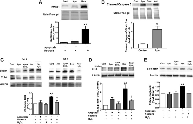
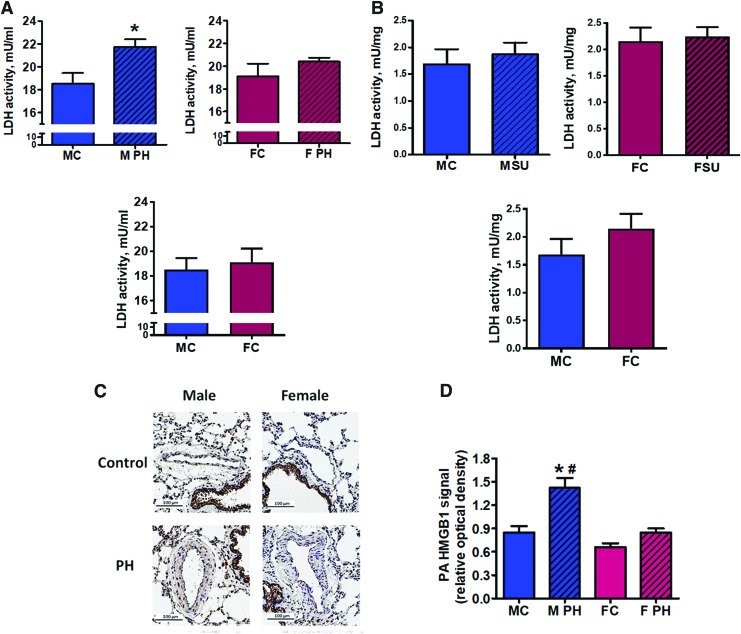
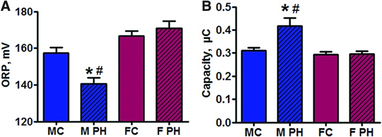
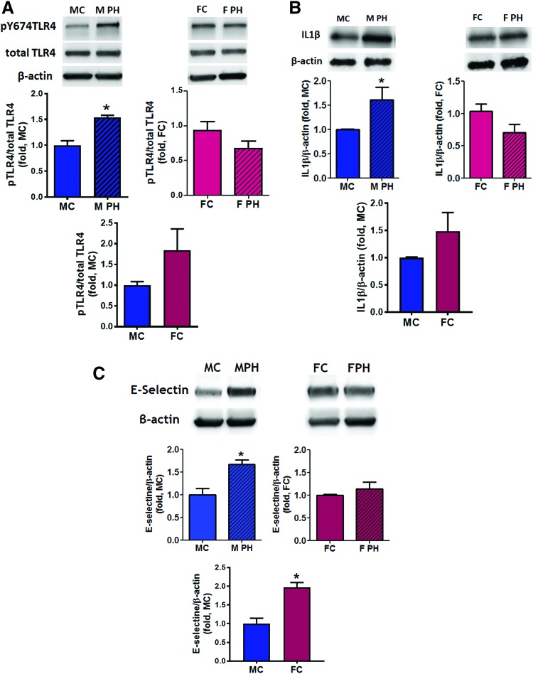
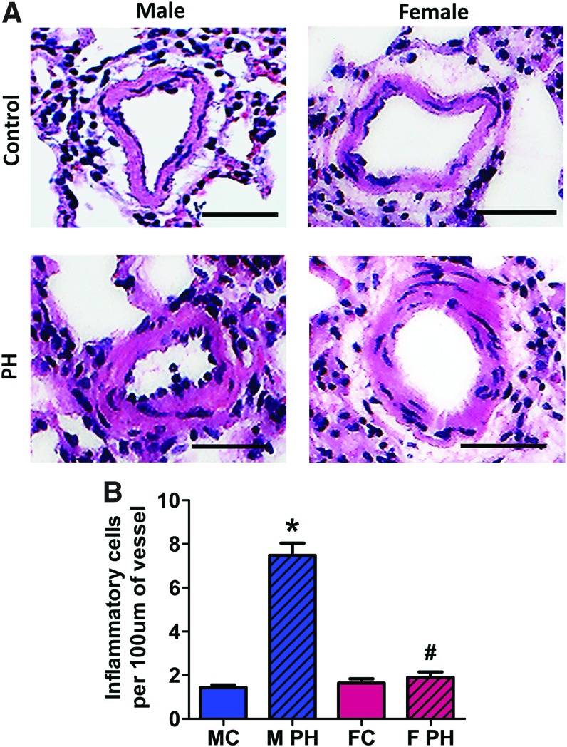
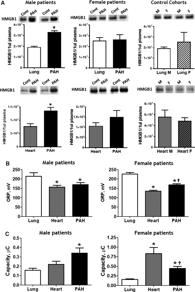
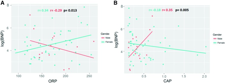
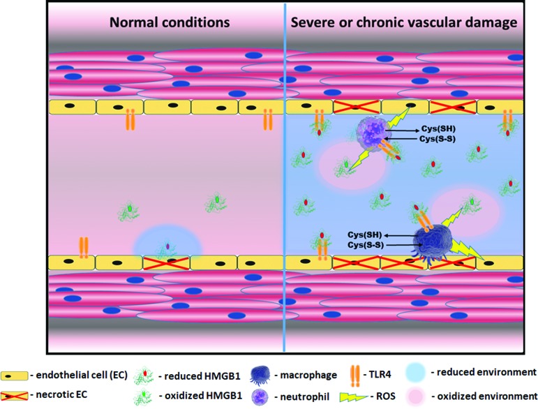
Aims: Pulmonary arterial hypertension (PAH) is a progressive lethal disease with a known gender dimorphism. Female patients are more susceptible to PAH, whereas male patients have a lower survival rate. Initial pulmonary vascular damage plays an important role in PAH pathogenesis. Therefore, this study aimed at investigating the role of gender in activation of apoptosis/necrosis-mediated signaling pathways in PAH. Results: The media collected from pulmonary artery endothelial cells (PAECs) that died by necrosis or apoptosis were used to treat naive PAECs. Necrotic cell death stimulated phosphorylation of toll-like receptor 4, accumulation of interleukin 1 beta, and expression of E-selectin in a redox-dependent manner; apoptosis did not induce any of these effects. In the animal model of severe PAH, the necrotic marker, high mobility group box 1 (HMGB1), was visualized in the pulmonary vascular wall of male but not female rats. This vascular necrosis was associated with male-specific redox changes in plasma, activation of the same inflammatory signaling pathway seen in response to necrosis in vitro, and an increased endothelial-leukocyte adhesion in small pulmonary arteries. In PAH patients, gender-specific changes in redox homeostasis correlated with the prognostic marker, B-type natriuretic peptide. Males had also shown elevated circulating levels of HMGB1 and pro-inflammatory changes. Innovation: This study discovered the role of gender in the initiation of damage-associated signaling in PAH and highlights the importance of the gender-specific approach in PAH therapy. Conclusion: In PAH, the necrotic cell death is augmented in male patients compared with female patients. Factors released from necrotic cells could alter redox homeostasis and stimulate inflammatory signaling pathways.

Keywords: gender difference; inflammation; necrosis; pulmonary hypertension.


Conflict of interest statement

No competing financial interests exist.

Figures

<b>FIG. 1.</b>
FIG. 1.
Reduced factors released from necrotic cells induce pro-inflammatory signaling and activation of intact endothelial cells. (A, B) Apoptosis was induced by incubation of PAEC in 2% of oxygen for 24 h, necrosis—by few free/thaw cycles. Necrosis but not apoptosis induced an accumulation of HMGB1 in cell culture media (A). Apoptosis induced Caspase 3 cleavage (B). The conditioned media collected from apoptotic or necrotic PAEC were used to treat intact PAEC. Factors released from necrotic, but not apoptotic cells induce phosphorylation of TLR4 (C), increase production of IL1β (D) and expression of E-selectin (E) in intact PAEC. The effects were attenuated when necrotic media were pretreated with H2O2 to induce oxidation of factors released from necrotic cells. Pretreatment with H2O2 did not induce any significant changes in PAEC treated by apoptotic media. Data are mean ± SEM, N = 3 per group (A); N = 4 per group (BE).*p < 0.05 versus Control; #p < 0.05 versus apoptotic cells or media; p < 0.05 versus reduced necrotic media in one-way ANOVA. ANOVA, analysis of variance; H2O2, hydrogen peroxide; HMGB1, high mobility group box 1; IL1β, interleukin 1 beta; PAEC, pulmonary artery endothelial cell; TLR4, toll-like receptor 4; SEM, standard error of the mean.
<b>FIG. 2.</b>
FIG. 2.
An increased level of necrosis is associated with pulmonary hypertension and male gender. Pulmonary hypertension induced an increase in the plasma level of LDH activity as a marker of tissue necrosis in male but not female rats (A) top left graph—males; top right graph—females; bottom graph—healthy controls; LDH activity in lungs show no difference in either gender compared with controls (B) top left graph—males; top right graph—females; bottom graph—healthy controls. Data are mean ± SEM, N = 5–7 per group. *p < 0.05 versus male control rats. p Value calculated by using unpaired t-test. The signal from extra-nuclear (non-co-localized with nuclei) HMGB1, an established marker of necrotic cell death, was visualized (C) and quantified (D) in the vascular wall of pulmonary arteries in Control and PAH male and female rats. Although PAH induced a strong PA vascular remodeling, a significant accumulation of extra-nuclear signal from HMGB1 was evidenced in male rats only (D). Data are mean ± SEM, N = 9–11 PA were analyzed for each rat with N = 6 rats in each group. *p < 0.05 versus male control rats; #p < 0.05 versus female with PAH rats in one-way ANOVA. MC indicates male control group; M PH—male PH group; FC—female control group; F PH—female PH group; LDH, lactate dehydrogenase; PA, pulmonary artery; PAH, pulmonary arterial hypertension; PH, pulmonary hypertension. Color images are available online.
<b>FIG. 3.</b>
FIG. 3.
Pulmonary hypertension-induced reductive shift is selectively associated with male gender. Two primary redox parameters—ORP (A) and Cap (B)—were measured in the plasma of control rats and rats with PAH. In male rats, a PAH-induced reductive shift in plasma was confirmed by decreased ORP and increased Cap. Data are mean ± SEM. (A) N = 12 for MC and M PH groups; N = 10 for FC; N = 8 for F PH; (B) N = 10 for MC, M PH, and FC; N = 8 for F PH. *p < 0.05 versus male control rats; #p < 0.05 versus female with PAH rats in one-way ANOVA. MC indicates male control group; M PH—male PH group; FC—female control group; F PH—female PH group. Cap, total antioxidant capacity; ORP, oxidation–reduction potential. Color images are available online.
<b>FIG. 4.</b>
FIG. 4.
Activation of pro-inflammatory signaling in male rats with PAH. In male, but not female rats, PAH development is associated with an activation of pro-inflammatory signaling and endothelial cells activation. The pulmonary tissue of male and female control rats and rats with PAH was used to measure the level of TLR4 phosphorylation as a marker of TLR4 activation (A) top left graph—males; top right graph—females; bottom graph—healthy controls, accumulation of pro-inflammatory cytokine IL1β (B) top left graph—males; top right graph—females; bottom graph—healthy controls and a protein level of E-selectin as a measure of pulmonary endothelial cell activation by cytokines (C) top left graph—males; top right graph—females; bottom graph—healthy controls. For all three measurements, there was a significant increase in pro-inflammatory signaling only in males with PAH; whereas in females, all pro-inflammatory markers were found to be comparable to the levels seen in control nonhypertensive rats. Data are mean ± SEM; (A, B) N = 4 for all groups; (C) N = 4 for MC, M PH, and FC; N = 5 for F PH group. *p < 0.05 versus male control rats. p Value calculated by using unpaired t-test. MC indicates male control group; M PH—male PH group; FC—female control group; F PH—female PH group. Color images are available online.
<b>FIG. 5.</b>
FIG. 5.
Pulmonary hypertension mediates leukocyte–endothelial cells adhesion in small pulmonary arteries of male rats. An increased level of endothelial–leukocyte adhesion molecule (E-selectin) expression in male PAH group correlated with an increase in the actual adhesion of leukocytes to endothelial layers in small PA of male rats. No increase in the leukocyte–endothelial interaction was found in female rats. (A) Representative images for each group (Control and PAH of both genders), (B) Quantification of leukocytes adhered to the endothelium of small PA. Data are mean ± SEM, N = 40 per group. *p < 0.05 versus male control rats; #p < 0.05 versus female with PAH rats in one-way ANOVA. Marker corresponds to 50 μM. MC indicates male control group; M PH—male PH group; FC—female control group; F PH—female PH group. Color images are available online.
<b>FIG. 6.</b>
FIG. 6.
Male patients with PAH have elevated levels of circulating HMGB1 and reductive stress. The HMGB1 signal produced by 1 μL of plasma was quantified by using Western blot analysis and compared within each gender between PAH and lung cohorts [(A), three upper panels] and PAH and heart cohorts [(A), three lower panels]. Only male PAH patients had increased HMGB1 plasma levels compared with control cohorts. Data are mean ± SEM. Upper panelN = 4 for lung male group; N = 6 for PAH male group; N = 10 for lung female group; N = 8 for PAH female group; lower panelN = 7 for all groups. *p < 0.05 versus lung or heart cohort. p Value calculated by using unpaired t-test. ORP (B) and Cap (C) were measured in the plasma samples of control cohorts and patients with PAH. Patients with non-PAH lung diseases showed evidence of strong oxidative stress, whereas patients with non-PAH heart diseases had reductive changes. Females with PAH had more oxidized redox parameters compared with the heart cohort, whereas male patients with PAH possessed equal (ORP) or even more reduced (Cap) plasma redox changes. Data are mean ± SEM, (B, C) N = 5 for lung; N = 8 for heart; N = 14 for PAH in male groups; N = 16 for lung; N = 9 for heart; N = 51 for PAH in female groups. *p < 0.05 versus lung cohort, p < 0.05 versus heart cohort. p Value calculated by using one-way ANOVA (Bonferroni's multiple-comparison test).
<b>FIG. 7.</b>
FIG. 7.
PAH patients show a gender difference in correlation between plasma redox parameters and markers of right ventricle dysfunction. Scatter plots showing the correlation between ORP—(A) and Cap—(B) and a biomarker of cardiac failure, BNP, in PAH patients. Adjustments are made to gender, age, and PAH therapy. The analysis indicates that the plasma redox environment significantly impacts BNP in PAH patients (p < 0.1). Moreover, this impact is significantly different with the interaction of genders (Pearson coefficients for ORP r = 0.34 [females] and r = −0.29 [males], p = 0.013, Pearson coefficients for Cap r = −0.18 [females] and r = 0.35 [males], p = 0.005). N = 50 for female PAH patients and N = 13 for male PAH patients. BNP, B-type natriuretic peptide. Color images are available online.
<b>FIG. 8.</b>
FIG. 8.
Gender difference in circulating WBC in patients with PAH. (A) PAH is associated with an increase in WBC compared with healthy individuals. The distribution of WBC values is shown as yellow columns. The dashed lines represent the mean WBC value. The solid lines show the mean WBC for the age-matched healthy individuals, as previously published (30). The inflammatory response is more profound in PAH male patients, whereas distribution in PAH females is more homogeneous and shows only a mild increase in WBC. (B) The levels of WBC in PAH male and female blood samples. N = 14 for male and N = 50 for female PAH patients. *p < 0.05 versus male PAH patients. p Value calculated by using unpaired t-test. WBC, white blood cell. Color images are available online.
<b>FIG. 9.</b>
FIG. 9.
Necrosis mediated activation of vascular inflammation. HMGB1 is active in its reduced state, whereas oxidation inhibits its activity. Therefore, the duration of HMGB1 signaling depends on the redox status of the extracellular environment, where it gets released on cell death. In a healthy state (left part of a scheme), even in a case of occasional necrosis, oxidized state of the extracellular environment ensures a quick inhibition of HMGB1. However, if the level of necrosis is relatively high like it was found in males, the local extracellular environment shifts to become more reduced due to the release of high amounts of intracellular glutathione and other thiols (right part of a scheme). This reduced environment prolongs HMGB1-mediated signaling. The reduced HMGB1 binds to a TLR4 receptor of endothelial cells and induces their activation. Active endothelial cells release cytokines, which attract macrophages and leukocytes to the site of damage and express the adhesive molecules that promote the recruitment of inflammatory cells. Activated macrophages and neutrophils, by producing ROS, oxidize HMGB1, but at the same time may additionally injure endothelial cells and, thus, promote damage-mediated inflammation. Moreover, activation of pattern recognition receptors such as TLR4 and RAGE induces further propagation of reductive stress by activation of cystine/cysteine redox cycle and secretion of large amounts of reduced free cysteine outside the cell (45). RAGE, receptor for advanced glycation end products; ROS, reactive oxygen species. Color images are available online.

Similar articles

Cited by

References

    1. Abe K, Toba M, Alzoubi A, Ito M, Fagan KA, Cool CD, Voelkel NF, McMurtry IF, and Oka M. Formation of plexiform lesions in experimental severe pulmonary arterial hypertension. Circulation 121: 2747–2754, 2010 - PubMed
    1. Aleshin VA, Artiukhov AV, Oppermann H, Kazantsev AV, Lukashev NV, and Bunik VI. Mitochondrial impairment may increase cellular NAD(P)H: resazurin oxidoreductase activity, perturbing the NAD(P)H-based viability assays. Cells 4: 427–451, 2015 - PMC - PubMed
    1. Amabile N, Heiss C, Chang V, Angeli FS, Damon L, Rame EJ, McGlothlin D, Grossman W, De Marco T, and Yeghiazarians Y. Increased CD62e(+) endothelial microparticle levels predict poor outcome in pulmonary hypertension patients. J Heart Lung Transplant 28: 1081–1086, 2009 - PubMed
    1. Bargagli E, Olivieri C, Bennett D, Prasse A, Muller-Quernheim J, and Rottoli P. Oxidative stress in the pathogenesis of diffuse lung diseases: a review. Respir Med 103: 1245–1256, 2009 - PubMed
    1. Basu S, Binder RJ, Suto R, Anderson KM, and Srivastava PK. Necrotic but not apoptotic cell death releases heat shock proteins, which deliver a partial maturation signal to dendritic cells and activate the NF-kappa B pathway. Int Immunol 12: 1539–1546, 2000 - PubMed

Publication types

MeSH terms

LinkOut - more resources