

Isolated Renal Mucormycosis in Immunocompetent Hosts: Clinical Spectrum and Management Approach
- PMID: 30652661
- PMCID: PMC6447097
- DOI: 10.4269/ajtmh.18-0103
Isolated Renal Mucormycosis in Immunocompetent Hosts: Clinical Spectrum and Management Approach
Abstract
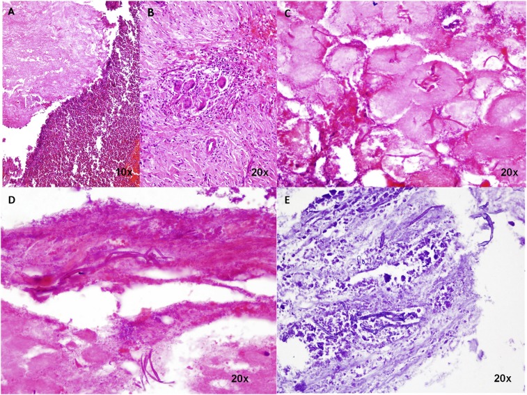
Isolated renal mucormycosis in immunocompetent hosts is a rare entity. We present the largest case series of isolated renal mucormycosis in immunocompetent hosts. Retrospective data of isolated renal mucormycosis from March 2012 to June 2017 was reviewed. Fifteen patients of isolated renal mucormycosis were identified. Contrast-enhanced computed tomography scan showed enlarged globular kidneys with decreased or patchy enhancement, perinephric stranding and thickened Gerota's fascia in all patients. Ten patients with unilateral involvement underwent nephrectomy and two of four patients with bilateral renal mucormycosis underwent bilateral nephrectomy. Two patients were managed with intravenous antifungal therapy alone. Overall, the mortality rate in our series was 40% (6/15). Isolated renal mucormycosis in healthy immunocompetent hosts is an emerging new entity. Prompt diagnosis based on the characteristic clinical and radiological picture and starting high-dose antifungal therapy at least 24 hours before surgical debridement offer the best chance of survival in these patients.
Figures


References
-
- Dhaliwal HS, et al. 2015Diagnosed only if considered: isolated renal mucormycosis. Lancet 385: 2322. - PubMed
-
- Pickles R, Long G, Murugasu R, 1994. Isolated renal mucormycosis. Med Journal Aust 160: 514–516. - PubMed
-
- Rogenes V, Vick S, Pultizer DR, 1998. Isolated renal mucormycosis. Infect Urol 113: 78–83.
MeSH terms
Substances
LinkOut - more resources
Full Text Sources
Medical
