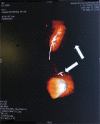
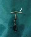
Migration of an intrauterine device causing severe hydronephrosis progressing to renal failure: A case report
- PMID: 30653092
- PMCID: PMC6370023
- DOI: 10.1097/MD.0000000000013872
Migration of an intrauterine device causing severe hydronephrosis progressing to renal failure: A case report
Abstract
Rationale: Intrauterine device (IUD) is commonly used in China. Its complications include uterine perforation, IUD ectopic migration, etc. However, a migrated IUD rarely leads to renal failure.
Patient concerns: IUD insertion in the patient was followed by unexplained pain in the left renal area, without bladder irritation or dysuresia.
Diagnoses: Hydronephrosis, renal failure, migrated IUD.
Interventions: The patient underwent laparoscopic and retroperitoneoscopic left nephrectomy, partial ureterectomy, and migrated IUD extraction.
Outcomes: No complications were found after 1 year of follow-up.
Lesson: An IUD should be placed by an experienced doctor. If conditions permit, it is best to perform the procedure under the guidance of ultrasound. The patients should be advised to undergo regular check-ups after the procedure. If necessary, abdominal color Doppler examination should be performed. Importantly, patients with IUD pregnancy must be reviewed.
Conflict of interest statement
The authors have no funding and conflicts of interest to disclose.
Figures


Similar articles
-
Hydronephrosis caused by intrauterine contraceptive device migration: three case reports with literature review.Clin Exp Obstet Gynecol. 2017;44(2):301-304. Clin Exp Obstet Gynecol. 2017. PMID: 29746046
-
Vesical transmigration of an intrauterine contraceptive device: A rare case report and literature review.Medicine (Baltimore). 2017 Oct;96(40):e8236. doi: 10.1097/MD.0000000000008236. Medicine (Baltimore). 2017. PMID: 28984781 Free PMC article. Review.
-
Emergent Laparoscopic Removal of a Perforating Intrauterine Device During Pregnancy Under Regional Anesthesia.J Minim Invasive Gynecol. 2019 Sep-Oct;26(6):1013-1014. doi: 10.1016/j.jmig.2019.03.012. Epub 2019 Mar 23. J Minim Invasive Gynecol. 2019. PMID: 30914327
-
Asymptomatic uterine perforation and IUD migration to the broad ligament: A case report.Medicine (Baltimore). 2024 Feb 16;103(7):e33857. doi: 10.1097/MD.0000000000033857. Medicine (Baltimore). 2024. PMID: 38363896 Free PMC article.
-
Sigmoid colon penetration by an intrauterine device: a case report and literature review.Mil Med. 2014 Jan;179(1):e127-9. doi: 10.7205/MILMED-D-13-00268. Mil Med. 2014. PMID: 24402999 Review.
Cited by
-
Intrauterine Contraceptive Device Migrated in the Urinary Tract: Case Report and Extensive Literature Review.J Clin Med. 2024 Jul 19;13(14):4233. doi: 10.3390/jcm13144233. J Clin Med. 2024. PMID: 39064273 Free PMC article.
-
Case report: An intrauterine device hugging the musculus rectus abdominis through the center of a cesarean scar.Front Surg. 2023 Jan 6;9:956856. doi: 10.3389/fsurg.2022.956856. eCollection 2022. Front Surg. 2023. PMID: 36684317 Free PMC article.
-
Spontaneous Urinoma Without Trauma or Obstruction in a 64-Year-Old Female.Cureus. 2020 Jul 17;12(7):e9241. doi: 10.7759/cureus.9241. Cureus. 2020. PMID: 32821587 Free PMC article.
-
Intrauterine device found in an ovarian tumor: A case report.Medicine (Baltimore). 2020 Oct 16;99(42):e22825. doi: 10.1097/MD.0000000000022825. Medicine (Baltimore). 2020. PMID: 33080762 Free PMC article.
References
-
- Harrison-Woolrych M, Ashton J, Coulter D. Uterine perforation on intrauterine device insertion: is the incidence higher than previously reported? Contraception 2003;67:53–6. - PubMed
-
- Zakin D, Stern WZ, Rosenblatt R. Complete and partial uterine perforation and embedding following insertion of intrauterine devices. II. Diagnostic methods, prevention, and management. Obstet Gynecol Surv 1981;36:401–17. - PubMed
-
- Shen Suqi, Li Ying Analysis of adverse events of ectopic iud in 90 cases. Chinese J Family Planning 2010;2:111–3.
-
- Tuncay YA, Tuncay E, Guzin K, et al. Transuterine migration as a complication of intrauterine contraceptive devices: six case reports. Eur J Contracept Reprod Health Care 2004;9:194–200. - PubMed
Publication types
MeSH terms
LinkOut - more resources
Full Text Sources

