

Characteristics and proposed classification system of posterior pilon fractures
- PMID: 30653144
- PMCID: PMC6370166
- DOI: 10.1097/MD.0000000000014133
Characteristics and proposed classification system of posterior pilon fractures
Abstract
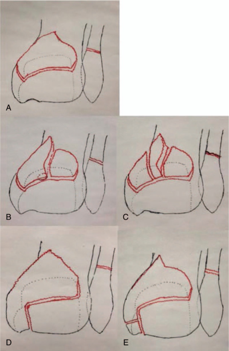
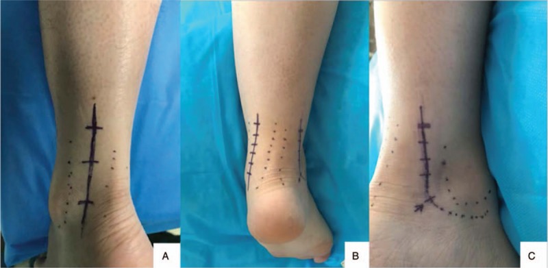
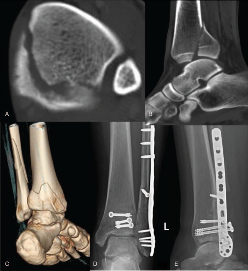
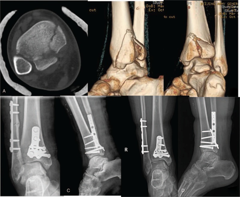
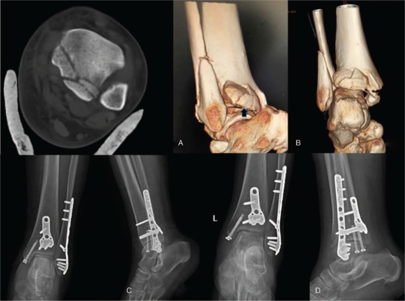
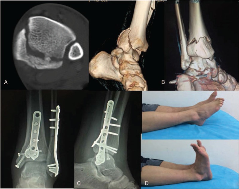
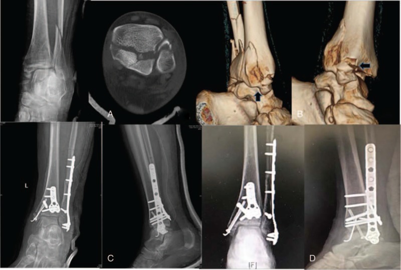
Posterior pilon fractures involve the medial malleolus (MM). Our purpose was to define the characteristics of posterior pilon fractures, and propose a classification system based on fracture morphology and type of management.The records of patients with posterior pilon fractures treated from 2011 to 2015 were retrospectively reviewed. The injury mechanism, fracture morphology, surgical approach, and follow-up results were reviewed and analyzed. This study was approved by the Institutional Review Board of PLA Army General Hospital.Thirty-six patients, 18 males and 18 females (mean age: 48.9 years) were included in the study. Four characteristics were used to define posterior pilon fractures. A simple posterolateral approach or a combined posterolateral and posteromedial approach was used for reduction and fixation in all patients. The mean follow-up time was 28.2 months, and at the end of follow-up, the mean American Orthopedic Foot and Ankle Society Score (AOFAS) was 82.5 points (range: 35-100 points). Based on injury mechanism and fracture morphology, we classified posterior pilon fractures into 3 types that suggest the optimal surgical approach: type I, a single complete fracture fragment; type II, a posterior malleolus fracture with 2 subtypes; type III, a posterior malleolus fracture associated with complete MM fracture with 2 subtypes.The proposed classification system based on injury mechanism and fracture morphology can guide the surgical approach to maximize outcomes.
Conflict of interest statement
The authors have no conflicts of interest to disclose.
Figures
Similar articles
-
[POSTEROLATERAL AND POSTEROMEDIAL APPROACHES FOR TREATMENT OF POSTERIOR Pilon FRACTURES IN ELDERLY PATIENTS].Zhongguo Xiu Fu Chong Jian Wai Ke Za Zhi. 2016 Sep 8;30(9):1089-1093. doi: 10.7507/1002-1892.20160222. Zhongguo Xiu Fu Chong Jian Wai Ke Za Zhi. 2016. PMID: 29786361 Chinese.
-
Fracture Line Morphology and a Novel Classification of Pilon Fractures.Orthop Surg. 2025 Feb;17(2):540-550. doi: 10.1111/os.14304. Epub 2024 Nov 23. Orthop Surg. 2025. PMID: 39579007 Free PMC article.
-
[Effectiveness of very low profile/variable angle locking plate internal fixation in treatment of posterior Pilon fractures extending to medial malleolus by posteromedial approach].Zhongguo Xiu Fu Chong Jian Wai Ke Za Zhi. 2014 May;28(5):558-61. Zhongguo Xiu Fu Chong Jian Wai Ke Za Zhi. 2014. PMID: 25073272 Chinese.
-
Complex Ankle Fractures: Practical Approach for Surgical Treatment.Foot Ankle Clin. 2020 Dec;25(4):587-595. doi: 10.1016/j.fcl.2020.08.002. Epub 2020 Sep 18. Foot Ankle Clin. 2020. PMID: 33543717 Review.
-
How to address the posterior malleolus in ankle fractures? A decision-making model based on the computerised tomography findings.Int Orthop. 2020 Jun;44(6):1177-1185. doi: 10.1007/s00264-020-04481-5. Epub 2020 Feb 4. Int Orthop. 2020. PMID: 32020283 Review.
Cited by
-
[Established classification systems of posterior malleolar fractures : A systematic literature review].Unfallchirurgie (Heidelb). 2023 May;126(5):387-398. doi: 10.1007/s00113-022-01162-3. Epub 2022 Apr 8. Unfallchirurgie (Heidelb). 2023. PMID: 35394158 Free PMC article. German.
-
Three-Dimensional Heat Map: The OTA/AO Type 43C Pilon Fracture Lines Distribution.Int J Gen Med. 2024 Jan 30;17:323-334. doi: 10.2147/IJGM.S444977. eCollection 2024. Int J Gen Med. 2024. PMID: 38314199 Free PMC article.
-
A systematic review of posterior pilon fractures.J Orthop. 2025 Jan 28;68:34-39. doi: 10.1016/j.jor.2025.01.034. eCollection 2025 Oct. J Orthop. 2025. PMID: 39995545 Review.
-
A systematic review of posterior pilon variant fractures.J Orthop. 2024 Feb 28;53:73-81. doi: 10.1016/j.jor.2024.02.035. eCollection 2024 Jul. J Orthop. 2024. PMID: 38476677 Free PMC article. Review.
-
Treatment outcomes of posterior pilon fractures using a simple single lateral approach via stretching fibular fracture line.Front Surg. 2023 Mar 30;10:1141606. doi: 10.3389/fsurg.2023.1141606. eCollection 2023. Front Surg. 2023. PMID: 37066001 Free PMC article.
References
-
- Chen DW, Li B, Yang YF, et al. Posterior pilon fractures. Foot Ankle Int 2013;34:766–7. - PubMed
-
- Topliss CJ, Jackson M, Atkins RM. Anatomy of pilon fractures of the distal tibia. J Bone Joint Surg Br 2005;87:692–7. - PubMed
-
- Huber M, Stutz PM, Gerber C. Open reduction and internal fixation of the posterior malleolus with a posterior antiglide plate using a postero-lateral approach— a preliminary report. Foot Ankle Surg 1996;2:95–103.
-
- Hansen S. Functional Reconstruction of the Foot and Ankle. 2000;Philadelphia PA: Lippincott Williams &Wilkins, 37–46.
-
- Amorosa LF, Brown GD, Greisberg JA. Surgical approach to posterior pilon fractures. J Orthop Trauma 2010;24:188–93. - PubMed
Publication types
MeSH terms
LinkOut - more resources
Full Text Sources
Medical
