

A computer-aided diagnosis system using artificial intelligence for the diagnosis and characterization of breast masses on ultrasound: Added value for the inexperienced breast radiologist
- PMID: 30653149
- PMCID: PMC6370030
- DOI: 10.1097/MD.0000000000014146
A computer-aided diagnosis system using artificial intelligence for the diagnosis and characterization of breast masses on ultrasound: Added value for the inexperienced breast radiologist
Abstract
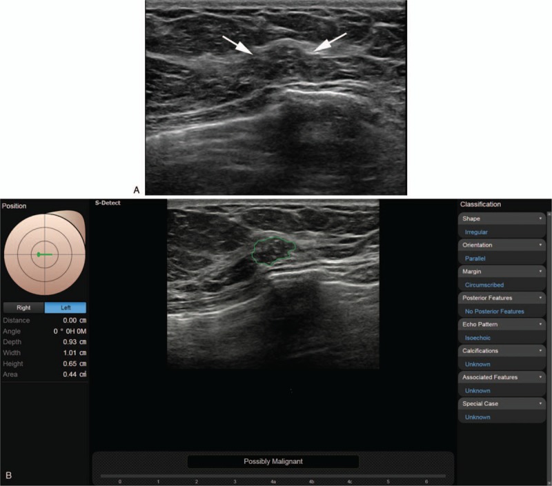
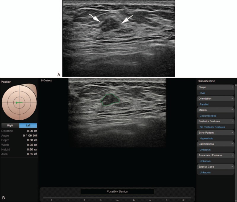
To evaluate the value of the computer-aided diagnosis (CAD) program applied to diagnostic breast ultrasonography (US) based on operator experience.US images of 100 breast masses from 91 women over 2 months (from May to June 2015) were collected and retrospectively analyzed. Three less experienced and 2 experienced breast imaging radiologists analyzed the US features of the breast masses without and with CAD according to the Breast Imaging Reporting and Data System (BI-RADS) lexicon and categories. We then compared the diagnostic performance between the experienced and less experienced radiologists and analyzed the interobserver agreement among the radiologists.Of the 100 breast masses, 41 (41%) were malignant and 59 (59%) were benign. Compared with the experienced radiologists, the less experienced radiologists had significantly improved negative predictive value (86.7%-94.7% vs 53.3%-76.2%, respectively) and area under receiver operating characteristics curve (0.823-0.839 vs 0.623-0.759, respectively) with CAD assistance (all P < .05). In contrast, experienced radiologists had significantly improved specificity (52.5% and 54.2% vs 66.1% and 66.1%) and positive predictive value (55.6% and 58.5% vs 64.9% and 64.9%, respectively) with CAD assistance (all P < .05). Interobserver variability of US features and final assessment by categories were significantly improved and moderate agreement was seen in the final assessment after CAD combination regardless of the radiologist's experience.CAD is a useful additional diagnostic tool for breast US in all radiologists, with benefits differing depending on the radiologist's level of experience. In this study, CAD improved the interobserver agreement and showed acceptable agreement in the characterization of breast masses.
Conflict of interest statement
The authors report no conflicts of interest.
Figures
References
-
- D’Orsi CJ. ACR BI-RADS Atlas: Breast Imaging Reporting and Data System. 5th ed.Reston: American College of Radiology; 2013.
-
- Doi K, MacMahon H, Katsuragawa S, Nishikawa RM, Jiang Y. Computer-aided diagnosis in radiology: potential and pitfalls. Eur J Radiol 1999;31:97–109. - PubMed
-
- Vyborny CJ, Giger ML, Nishikawa RM. Computer-aided detection and diagnosis of breast cancer. Radiol Clin North Am 2000;38:725–40. - PubMed
-
- Giger ML, Karssemeijer N, Armato SG., 3rd Computer-aided diagnosis in medical imaging. IEEE Trans Med Imaging 2001;20:1205–8. - PubMed
MeSH terms
LinkOut - more resources
Full Text Sources
Medical
Miscellaneous
