. 2019 Jan 17;13(1):e0007108.
doi: 10.1371/journal.pntd.0007108. eCollection 2019 Jan.

Development of a preliminary in vitro drug screening assay based on a newly established culturing system for pre-adult fifth-stage Onchocerca volvulus worms

Affiliations

Development of a preliminary in vitro drug screening assay based on a newly established culturing system for pre-adult fifth-stage Onchocerca volvulus worms

Denis Voronin et al. PLoS Negl Trop Dis. .

Abstract

Background: The human filarial parasite Onchocerca volvulus is the causative agent of onchocerciasis (river blindness). It causes blindness in 270,000 individuals with an additional 6.5 million suffering from severe skin pathologies. Current international control programs focus on the reduction of microfilaridermia by annually administering ivermectin for more than 20 years with the ultimate goal of blocking of transmission. The adult worms of O. volvulus can live within nodules for over 15 years and actively release microfilariae for the majority of their lifespan. Therefore, protracted treatment courses of ivermectin are required to block transmission and eventually eliminate the disease. To shorten the time to elimination of this disease, drugs that successfully target macrofilariae (adult parasites) are needed. Unfortunately, there is no small animal model for the infection that could be used for discovery and screening of drugs against adult O. volvulus parasites. Here, we present an in vitro culturing system that supports the growth and development of O. volvulus young adult worms from the third-stage (L3) infective stage.

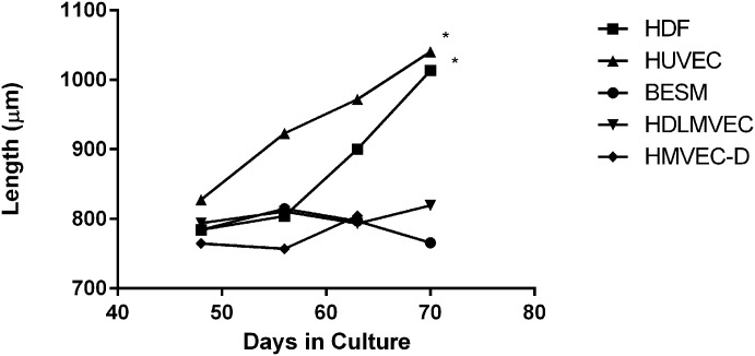
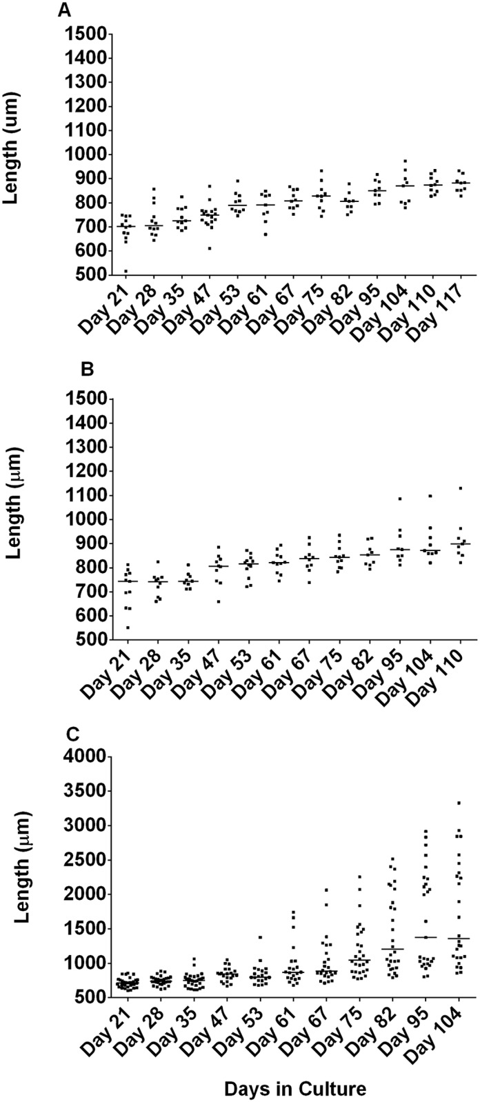
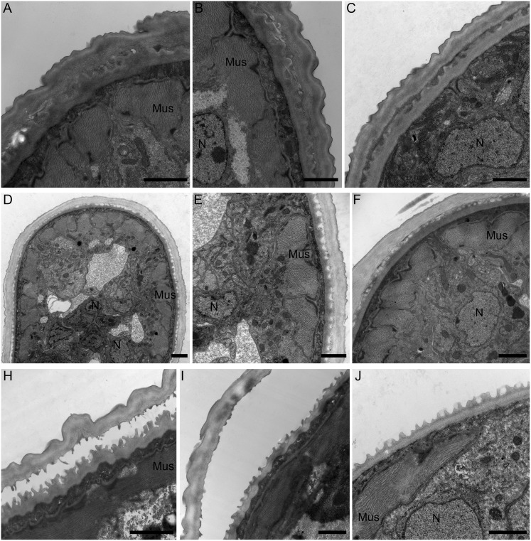
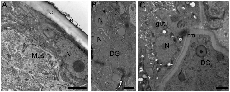
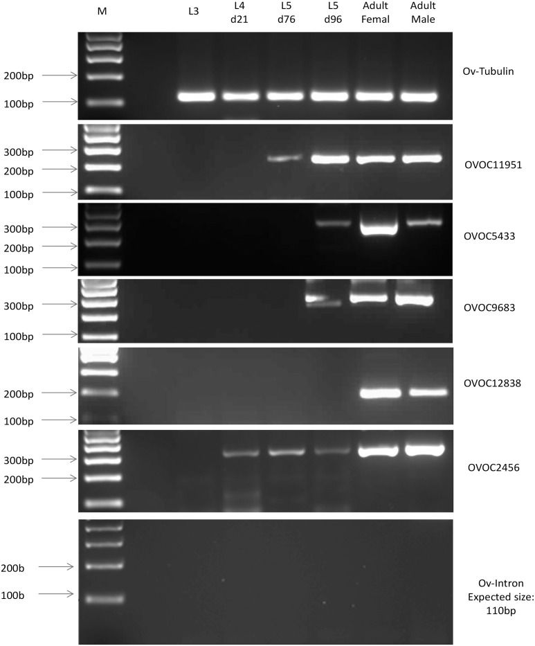
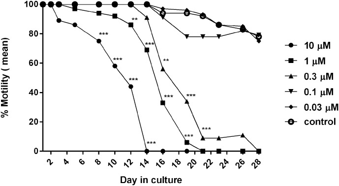
Methodology/principal findings: In this study we optimized the culturing system by testing several monolayer cell lines to support worm growth and development. We have shown that the optimized culturing system allows for the growth of the L3 worms to L5 and that the L5 mature into young adult worms. Moreover, these young O. volvulus worms were used in preliminary assays to test putative macrofilaricidal drugs and FDA-approved repurposed drugs.

Conclusion: The culture system we have established for O. volvulus young adult worms offers a promising new platform to advance drug discovery against the human filarial parasite, O. volvulus and thus supports the continuous pursuit for effective macrofilaricidal drugs. However, this in vitro culturing system will have to be further validated for reproducibility before it can be rolled out as a drug screen for decision making in macrofilaricide drug development programs.


Conflict of interest statement

Cell Systems- 3D, LLC is a research for-profit contract research organization (CRO) whose focus is the development of 3-D in vitro models of human, veterinary or insect cells/tissues as investigatory platforms for development of successful countermeasures of infectious diseases and/or agents. MTS was the sole employee of Cell Systems-3D, LLC involved in this research effort. The company participation in Cell Systems-3D,LLC nor any current or past employee has a commercial or employment affiliation with the New York Blood Bank or the Bill and Melinda Gates Foundation. The Company participation in this publication is as a research co-investigator. MTS is the founder and Chief Science Officer of the company.

Figures

Fig 1
Fig 1. Dynamic OvL4-L5 growth in 96-well plate settings.
HDF and HUVEC showed the best support for worms’ growth (day 70: grown on the HDF monolayers—range of 803–1374 μm, p = 0.03 (n = 7) as compared to BESM; grown on HUVEC monolayers—range of 804–1504 μm, p = 0.03 (n = 7) as compared to BESM). L4 cultured in HMVEC-D, BESM or HDLMVEC had the lowest growth rate (ANOVA test p = 0.016 (n = 9), d56) and highest mortality. *—p<0.05.
Fig 2
Fig 2. Comparing growth of OvL4 to OvL5 in 24-well plate containing a monolayer of HUVEC and in the presence of varying culture media composition.
A. OvL4-media with 20% FBS; B. OvL4-media with 20% FBS and 0.1% lipid mixture (OvL4-CM); C. OvL4-media with 25% FBS, 0.1% lipid mixture, and 1% mix of amino acids (OvL4-CMS). OvL5 cultured in OvL4-CMS media had the longest length (on day 104 –range of 857–3329 μm, median 1358 μm; p = 0.002 (n = 26) as compared to the length of worms in OvL4-CM media with 20% FBS (A), p = 0.003 (n = 26) as compared to the length of worms in OvL4-CM media with 20% FBS and 0.1% lipid mixture (B)).
Fig 3
Fig 3. Transmission electron microscopy of OvL4 during molting to OvL5.
A-C, Growth of new cuticle (c) beneath the old one (arrowheads) (day 48–50); D-F, beginning of cuticle separation (worms on day 50–60); H-J, final stage of cuticle separation (worms on days 60–75). Worms also had well-developed muscle filaments (Mus), and normal morphology of cells. Abbreviation: N—nuclei. Bar: A-C– 1 μm; D– 2 μm; E-J– 1 μm.
Fig 4
Fig 4. Microphotographs of O. volvulus (120 days in culture) show normal morphology of cells and developing organs.
A, worms have a cuticle and epicuticle layer, also showed normal organization of muscle filaments (Mus). B, C, young adults have germ cells organized in developing gonads (DG) with basal membrane (bm). Abbreviation: N—nuclei. Bar A, C– 1 μm; B– 2 μm.
Fig 5
Fig 5. Gel electrophoresis of PCR products obtained using cDNA prepared from L3, L4 D21, L5 D76, L5 D96, adult males and females, and primers corresponding to selected stage-specific biomarkers (S1 Table).
Tubulin was used as quality control for cDNA and PCR. Primers for an intron (expected product size– 110 bp) were used as a negative control to test that there was no contamination of genomic DNA. M—Marker.
Fig 6
Fig 6. Motility of O. volvulus L5 worms exposed to different concentrations of flubendazole.
Treatment was for 14 days with an additional 14 day follow up without the drug. Flubendazole (0.3 μM and higher) caused significant inhibition of worms’ motility. Specifically: 10 μM of the drug caused significant (***—p<0.001, n = 9) inhibition of motility as compared with control (n = 14) starting on the 9th day; 1 μM of the drug caused significant (**—p<0.01, n = 11) inhibition of motility as compared with control (n = 14) starting on the 12th day; and 0.3 μM of drug caused significant (p<0.01, n = 8) inhibition of motility as compared with control (n = 14) starting on the 16th day. Motility was observed using a microscope and scored based on the following scale: 100% motility, constant coiling movement; 75% motility, slower coiling; 50% motility, slow and intermittent movement; 25% motility, very slow movement or twitching; and 0% motility, no movement.

References

    1. WHO [Internet]. http://www.who.int/mediacentre/factsheets/fs095/en/
    1. Coffeng LE, Stolk WA, Zouré HGM, Veerman JL, Agblewonu KB, Murdoch ME, et al. African Programme for Onchocerciasis Control 1995–2015: Updated Health Impact Estimates Based on New Disability Weights. PLoS Negl Trop Dis. 2014;8 10.1371/journal.pntd.0002759 - DOI - PMC - PubMed
    1. Lustigman S, Huima T, Brotman B, Miller K, Prince AM. Onchocerca volvulus: biochemical and morphological characteristics of the surface of third- and fourth-stage larvae. Exp Parasitol. 1990;71: 489–495. Available: http://www.ncbi.nlm.nih.gov/entrez/query.fcgi?cmd=Retrieve&db=PubMed&dop... - PubMed
    1. Morris CP, Evans H, Larsen SE, Mitre E. A Comprehensive, model-based review of vaccine and repeat infection trials for filariasis. Clin Microbiol Rev. 2013;26: 381–421. 10.1128/CMR.00002-13 - DOI - PMC - PubMed
    1. Abraham D, Lange AM, Yutanawiboonchai W, Trpis M, Dickerson JW, Swenson B, et al. Survival and development of larval Onchocerca volvulus in diffusion chambers implanted in primate and rodent hosts. J Parasitol. 1993;79: 571–582. 10.2307/3283385 - DOI - PubMed

Publication types