. 2019 Jan 17;15(1):e1007891.
doi: 10.1371/journal.pgen.1007891. eCollection 2019 Jan.

Single-molecule dynamics and genome-wide transcriptomics reveal that NF-kB (p65)-DNA binding times can be decoupled from transcriptional activation

Affiliations

Single-molecule dynamics and genome-wide transcriptomics reveal that NF-kB (p65)-DNA binding times can be decoupled from transcriptional activation

Andrea Callegari et al. PLoS Genet. .

Abstract

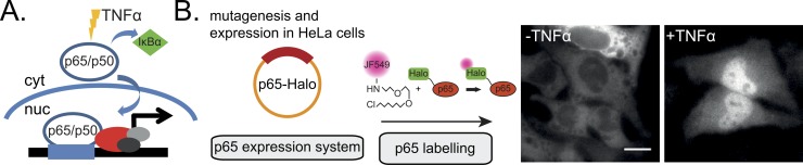
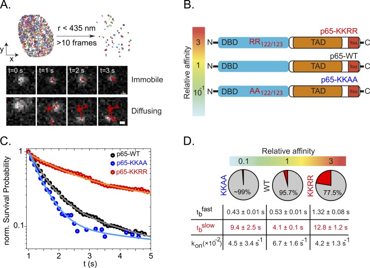
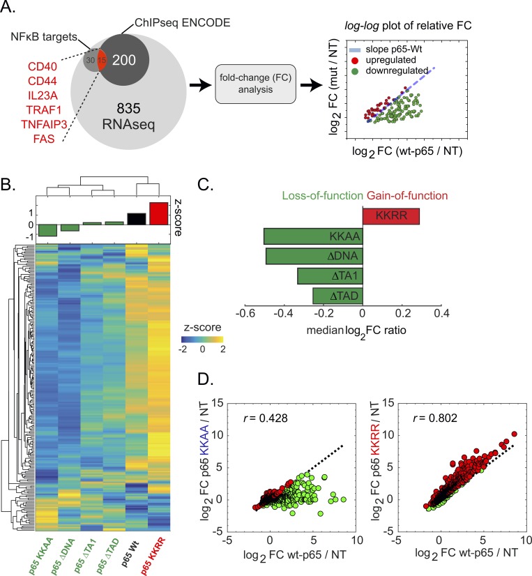
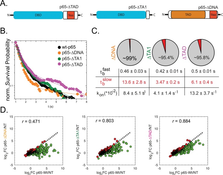
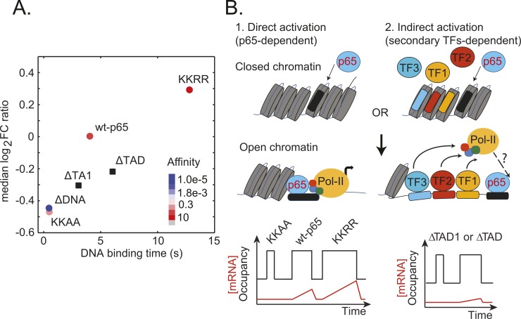
Transcription factors (TFs) regulate gene expression in both prokaryotes and eukaryotes by recognizing and binding to specific DNA promoter sequences. In higher eukaryotes, it remains unclear how the duration of TF binding to DNA relates to downstream transcriptional output. Here, we address this question for the transcriptional activator NF-κB (p65), by live-cell single molecule imaging of TF-DNA binding kinetics and genome-wide quantification of p65-mediated transcription. We used mutants of p65, perturbing either the DNA binding domain (DBD) or the protein-protein transactivation domain (TAD). We found that p65-DNA binding time was predominantly determined by its DBD and directly correlated with its transcriptional output as long as the TAD is intact. Surprisingly, mutation or deletion of the TAD did not modify p65-DNA binding stability, suggesting that the p65 TAD generally contributes neither to the assembly of an "enhanceosome," nor to the active removal of p65 from putative specific binding sites. However, TAD removal did reduce p65-mediated transcriptional activation, indicating that protein-protein interactions act to translate the long-lived p65-DNA binding into productive transcription.


Conflict of interest statement

The authors have declared that no competing interests exist.

Figures

Fig 1
Fig 1. NFκB-p65 mode of action and experimental setup.
(A) p65 forms a heterodimeric complex with p50 in the cytosol (cyt). The complex is bound by the cytosolic inhibitor IκBa that prevents its translocation into the nucleus. Following TNF-α stimulation and IκBα dissociation, p65/p50 translocate into the nucleus allowing subsequent DNA binding and gene activation. (B) We used a copy of the human p65 fused to a Halo tag as a basis for our mutational expression system. P65-Halo constructs were then expressed in HeLa cells and fluorescently labelled with a JF549 Halo ligand. Upon TNF-α stimulation, labelled p65-Halo translocates into the nucleus. Scale bar: 10 μm.
Fig 2
Fig 2. DNA binding time of p65 DNA affinity mutants.
(A) Single particle tracking (SPT) was performed after TNF-α stimulation and p65-Halo translocation into the nucleus. All recorded trajectories were filtered based on a spatial threshold established using an immobile control (Histone subunit H2B, see S3 Fig). After filtering out mobile molecules (i) and correction of photobleaching, the DNA binding time could be estimated from the length of each individual trajectory. (B) Schematic overview of p65 affinity mutants. (C) Normalized survival probability plots (1-CDF plot) of the DNA-bound fraction for p65-WT as well as the DNA affinity mutants KKAA and KKRR. The distributions were fitted using a bi-exponential function revealing the fast (tbfast) and slow (tbslow) DNA binding times. (D) Summary of the obtained fitting parameters together with the relative DNA dissociation constant KD for each construct. The pie chart shows the fraction of events associated to the fast (grey) or slow binding time. Kon* was obtained from single step displacement histograms as described in Methods. While all constructs exhibit similar kon* as well as tbfast, the slow binding time tbslow correlates with the DNA affinity.
Fig 3
Fig 3. Transcriptional activity of p65 DNA affinity mutants.
(A) We assessed the level of gene activation using RNA sequencing (RNA-Seq). To this end, total mRNA was isolated and sequenced using next-generation sequencing. The total initial set of 1080 genes was cross-referenced using the Chip-Seq ENCODE database, providing a subset of 215 direct interacting genes. A second subset of 45 genes consist of known NFκB regulated genes. For each p65 mutant, the fold-change (FC) expression above the non-transfected (NT) control was calculated and for each gene compared with p65-WT using a log-log FC plot. As a general discrimination between up- and downregulated genes compared to p65-WT, we calculated the logFC ratio. Values with logFC ratio>1 are marked upregulated (red), those with logFC ratio<1 are marked downregulated (green). (B) FC values were standardized (per gene) and the different conditions clustered hierarchically to identify similarities. Interestingly, p65-WT co-clusters with p65-KKRR, which also shows the highest average z-score (B, top plot) and was identified as the only gain-of-function mutant (C). The two transactivation mutants as well as the low affinity mutant and p65-ΔDNA also co-cluster highlighting their functional similarity. (C) Classification of each p65 variant based on the logFC ratio estimator, showing that p65-KKRR (i.e. with higher DNA affinity) represents the only gain-of-function mutant. (D) RNA-Seq analysis comparing transcriptional activation of p65-KKAA and p65-KKRR with p65-WT. p65-KKAA shows very weak correlation with p65-WT as well as a strongly reduced gene activation (logFC ratio = -0.47) indicating a loss of gene specificity as well as activation potential. In contrast, p65-KKRR shows higher correlation as well as an increased gene activation (logFC ratio = 0.28).
Fig 4
Fig 4. DNA binding and transcriptional activity of p65 truncation mutants.
(A) Schematic overview of p65 truncation mutants. (B) Normalized survival probability (1-CDF plot) plots of the DNA-bound fraction for p65-WT as well as the transactivation mutants ΔTA1 and ΔTAD as well as a mutant with removed DNA-binding domain (ΔDNA). The distributions were fitted using a bi-exponential function revealing the fast (tbfast) and slow (tbslow) DNA binding times. (C) As for the DNA affinity mutants, we found kon* to be in a similar range for all the tested constructs. (D) RNA-Seq analysis revealed very low residual transcriptional activation of p65-ΔDNA as evident by logFC ratio = -0.45. The two transactivation mutants showed good correlation with p65-WT (r ~ 0.8) but at strongly reduced transcript abundance resulting in logFC ratio around -0.25.
Fig 5
Fig 5. Correlation between p65 mutants’ transcriptional activity, DNA-affinity and binding time.
(A) The median log2 FC ratio as retrieved from RNA-Seq data is plotted against tbslow. Note that ΔDNA affinity has been assigned to an arbitrarily low value. (B) Working model for p65 mediated transcriptional activation. (1) P65 can act as a pioneering TF, open the chromatin and bind its consensus DNA sequence. Transcriptional activation can then be initiated although the exact mechanism of RNA pol-II recruitment remains unclear. Following this model, the DNA binding time would correlate with the transcriptional output, while removal of TADs would not affect the complex stability. (2) An important extension of this model as suggested by our data is that TADs are required to efficiently translate p65 DNA binding into transcriptional output presumably through the recruitment of protein co-factors.

References

    1. Medzhitov R, Horng T. Transcriptional control of the inflammatory response. Nat Rev Immunol. 2009;9: 692–703. 10.1038/nri2634 - DOI - PubMed
    1. Baeuerle PA, Baltimore D. IκB: A specific inhibitor of the NF-κB transcription factor. Science (80-). 1988;242: 540–546. - PubMed
    1. Baeuerle PA, Henkel T. Function and activation of NF-κB in the immune system. Annu Rev Immunol. 1994;12: 141–179. 10.1146/annurev.iy.12.040194.001041 - DOI - PubMed
    1. Müller CW, Van D, Sodeoka M, Verdine GL, Harrison SC. Structure of the nf-κb p50 homodimer bound to dna. Nature. 1995;373: 311–317. 10.1038/373311a0 - DOI - PubMed
    1. Ghosh G, Van D, Ghosh S, Sigler PB. Structure of nf-κb p50 homodimer bound to a κb site. Nature. 1995;373: 303–310. 10.1038/373303a0 - DOI - PubMed

Publication types

MeSH terms