. 2019 Jan 17;14(1):e0210889.
doi: 10.1371/journal.pone.0210889. eCollection 2019.

The effect of sodium butyrate and cisplatin on expression of EMT markers

Affiliations

The effect of sodium butyrate and cisplatin on expression of EMT markers

Alena Mrkvicova et al. PLoS One. .

Abstract

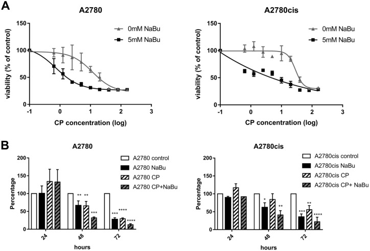
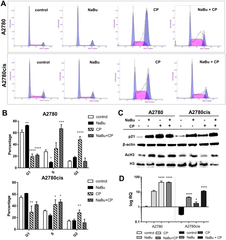
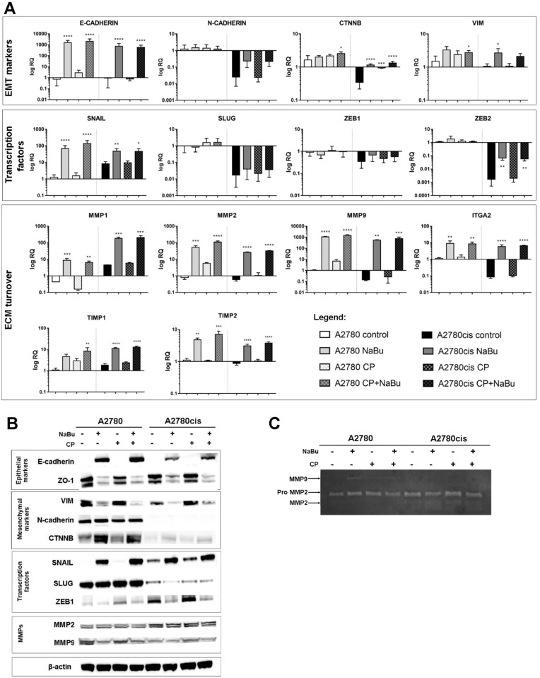
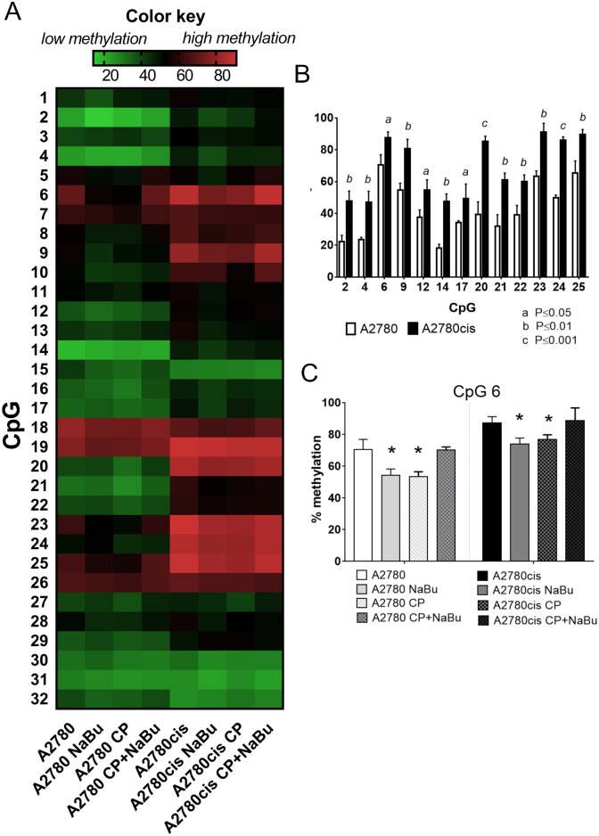
Histone modifications play a key role in the epigenetic regulation of gene transcription in cancer cells. Histone acetylations are regulated by two classes of enzymes, histone acetyltransferases (HATs) and histone deacetylases (HDACs). HDACs are increased in ovarian carcinomas and they are involved in carcinogenesis and resistance to chemotherapeutic agents. In our study we investigated anticancer effect of HDAC inhibitor sodium butyrate (NaBu) on cisplatin-sensitive and cisplatin-resistant ovarian cell lines A2780 and A2780cis. A2780 and A2780cis were treated with NaBu alone or in combination with cisplatin (CP). NaBu inhibited the growth of both cell lines and enhanced cytotoxic effect of CP. Exposure to NaBu for 24 h induced cell cycle arrest. The expressions of EMT-related genes and proteins were further investigated by qPCR and western blot analysis. Loss of E-cadherin has been shown to be crucial in ovarian cancer development. We found that NaBu dramatically induce expression of E-cadherin gene (CDH1) and protein levels in A2780 and A2780cis. We investigated correlation between transcription and methylation of CDH1gene. Methylation level analysis in 32 CpG sites in CDH1 gene (promoter/exon1 regions) was performed using bisulfite NGS (Next Generation Sequencing). We found that cisplatin-resistant cell line A2780cis cells differ from their cisplatin-sensitive counterparts in the CDH1 methylation. Methylation in A2780cis cells is elevated compared to A2780. However, NaBu-induced expression of CDH1 was not accompanied by CDH1 demethylation. NaBu treatment induced changes in expression of EMT-related genes and proteins. Interestingly E-cadherin zinc finger transcriptional repressor SNAIL1 was upregulated in both cell lines. Mesenchymal marker vimentin was downregulated. Matrix metalloproteases (MMPs) are necessary for pericellular proteolysis and facilitate migration and invasion of tumour cells. NaBu induced mRNA expression of MMPs, mild changes in activities of gelatinases MMP2 and MMP9 were detected. Our data demonstrate that NaBu sensitizes cisplatin-resistant ovarian cancer cells, re-established E-cadherin expression, but it was not able to reverse the EMT phenotype completely.


Conflict of interest statement

The authors have declared that no competing interests exist.

Figures

Fig 1
Fig 1. Sodium butyrate enhances the cytotoxic effect of cisplatin.
Cell proliferation/viability was measured using colorimetric WST-1 assay. Data are presented as the percentage relative to untreated cells, arbitrary set to 100%. Dose response of A2780 and A2780cis cells to 48 h treatment with CP alone or in combination with NaBu (5 mM) (A). Each point represents the mean ± SD from 2 independent experiments. Dynamic of antiproliferative effect of NaBu (5 mM) alone and in combination with CP (5 μM) (B). The data points in the graph are the means ± SD of 3 independent experiments. Significance was tested using one-way ANOVA. *P ≤ 0.05, **P ≤ 0.01, *** P≤ 0.001, **** P≤ 0.0001 are significantly different from control values.
Fig 2
Fig 2. Effect of cisplatin and butyrate on the cell cycle and histone acetylation.
Distribution of A2780 and A2780cis cells in G1, S, and G2/M phases of the cell cycle after 5 mM NaBu, 5 μM CP single-agent treatments and combination of both treatments was measured by flow cytometry (A, B). Panel A shows representative cell cycle analysis histograms. The bar graph (panel B) represents cumulative data on cell cycle distribution in the G1, S, and G2/M phase. The data points in the graph are the means ± SD of 3 independent experiments. Significance was tested using one-way ANOVA (*P ≤ 0.05, **P ≤ 0.01, *** P≤ 0.001, **** P≤ 0.0001 vs. control). p21, histone H3 and acetylated H3 protein amount was detected by immunoblot analysis (C). Blots shown are representative of at least three independent experiments. β-Actin was used as a loading control. Gene expression of CDKN1 in A2780 and A2780cis cells was measured using qPCR (D). The results were normalized for the HPRT expression. Data are presented as means ± SD of 3 independent experiments. Significance was tested using one-way ANOVA. (*P ≤ 0.05, **P ≤ 0.01, *** P≤ 0.001, **** P≤ 0.0001 vs. control).
Fig 3
Fig 3. EMT related genes and proteins expression.
Gene expression of EMT related genes in A2780 and A2780cis cells was measured using qPCR (A). Results were normalized for the HPRT expression. Data are presented as means ± SD of 3 independent experiments. Significance was tested using one-way ANOVA (*P ≤ 0.05, **P ≤ 0.01, *** P≤ 0.001, **** P≤ 0.0001 vs. control). Protein expression was detected by immunoblot analysis (B). A representative immunoblot of at least three independent experiments is presented. β-Actin was used as a loading control. Activities of MMP-2 and MMP-9 in the cell culture supernatants were measured by gelatin zymography. Gelatinolytic activity of pro and active MMP-2 and active MMP-9 are visible as a clear area on the gel (C).
Fig 4
Fig 4. CDH1 gene methylation.
Heat map displays changes in CDH1 methylation in 32 CpG sites in A2780 and A2780cis cells treated with NaBu, CP or combination of both (A). Analysis was performed in three independent experiments, means are shown. Significantly hypermethylated individual CpG sites in A2780cis cells compared to A2780 cells (B). The effect of NaBu, CP or combined treatment on percentage of methylation at CpG site 6 (C). The plots represent means ± SD of 3 independent experiments. Significance was tested using Student t-test P ≤ 0.05.

Similar articles

Cited by

References

    1. Reid BM, Permuth JB, Sellers TA. Epidemiology of ovarian cancer: a review. Cancer Biol Med. 2017;14(1):9–32. 10.20892/j.issn.2095-3941.2016.0084 - DOI - PMC - PubMed
    1. Curradi M, Izzo A, Badaracco G, Landsberger N. Molecular mechanisms of gene silencing mediated by DNA methylation. Mol Cell Biol. 2002;22(9):3157–73. 10.1128/MCB.22.9.3157-3173.2002 - DOI - PMC - PubMed
    1. Jin KL, Pak JH, Park JY, Choi WH, Lee JY, Kim JH, et al. Expression profile of histone deacetylases 1, 2 and 3 in ovarian cancer tissues. Journal of gynecologic oncology. 2008;19(3):185–90. 10.3802/jgo.2008.19.3.185 - DOI - PMC - PubMed
    1. Eckstein N. Platinum resistance in breast and ovarian cancer cell lines. J Exp Clin Canc Res. 2011;30 10.1186/1756-9966-30-91 - DOI - PMC - PubMed
    1. Koberle B, Tomicic MT, Usanova S, Kaina B. Cisplatin resistance: Preclinical findings and clinical implications. Bba-Rev Cancer. 2010;1806(2):172–82. 10.1016/j.bbcan.2010.07.004 - DOI - PubMed

Publication types

MeSH terms