. 2019 Mar;33(3):798-807.
doi: 10.1002/ptr.6273. Epub 2019 Jan 17.

HIF-1α/VEGF signaling-mediated epithelial-mesenchymal transition and angiogenesis is critically involved in anti-metastasis effect of luteolin in melanoma cells

Affiliations

HIF-1α/VEGF signaling-mediated epithelial-mesenchymal transition and angiogenesis is critically involved in anti-metastasis effect of luteolin in melanoma cells

Chunyu Li et al. Phytother Res. 2019 Mar.

Abstract

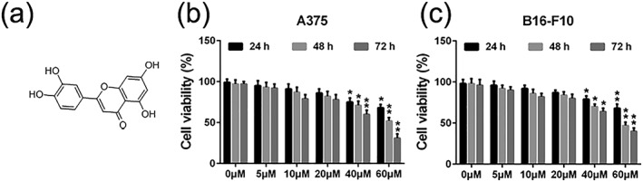
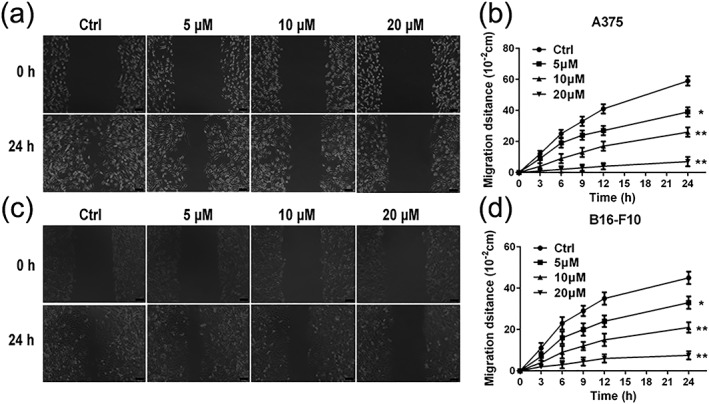
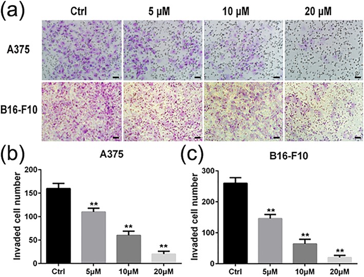
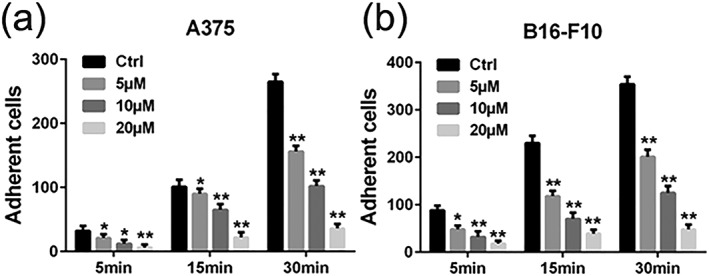
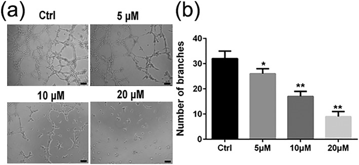
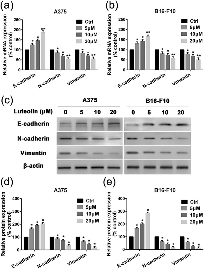
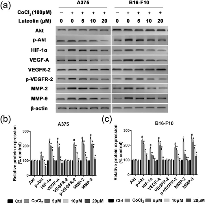
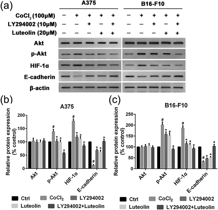
Tumor metastasis is still the leading cause of melanoma mortality. Luteolin, a natural flavonoid, is found in fruits, vegetables, and medicinal herbs. The pharmacological action and mechanism of luteolin on the metastasis of melanoma remain elusive. In this study, we investigated the effect of luteolin on A375 and B16-F10 cell viability, migration, invasion, adhesion, and tube formation of human umbilical vein endothelial cells. Epithelial-mesenchymal transition (EMT) markers and pivotal molecules in HIF-1α/VEGF signaling expression were analysed using western blot assays or quantitative real-time polymerase chain reaction. Results showed that luteolin inhibits cellular proliferation in A375 and B16-F10 melanoma cells in a time-dependent and concentration-dependent manner. Luteolin significantly inhibited the migratory, invasive, adhesive, and tube-forming potential of highly metastatic A375 and B16-F10 melanoma cells or human umbilical vein endothelial cells at sub-IC50 concentrations, where no significant cytotoxicity was observed. Luteolin effectively suppressed EMT by increased E-cadherin and decreased N-cadherin and vimentin expression both in mRNA and protein levels. Further, luteolin exerted its anti-metastasis activity through decreasing the p-Akt, HIF-1α, VEGF-A, p-VEGFR-2, MMP-2, and MMP-9 proteins expression. Overall, our findings first time suggests that HIF-1α/VEGF signaling-mediated EMT and angiogenesis is critically involved in anti-metastasis effect of luteolin as a potential therapeutic candidate for melanoma.

Keywords: HIF-1α/VEGF signaling; angiogenesis; epithelial-mesenchymal transition; luteolin; melanoma; metastasis.


Conflict of interest statement

The authors declare that they have no conflicts of interest.

Figures

Figure 1
Figure 1
Effects of luteolin on the viability of melanoma cells A375 and B16‐F10. (a) Chemical structure of luteolin. (b) A375 and (c) B16‐F10 cells were treated with indicated concentrations of luteolin (5, 10, 20, 40, and 60 μM) for 24, 48, and 72 hr, then cell viability was measured by MTT assay. Values are the means ± SD from three independent determinations. *p < 0.05 and **p < 0.01 indicate a significant difference from the control group
Figure 2
Figure 2
Effects of luteolin on the migratory ability of melanoma cells. (a and b) A375 and (c and d) B16‐F10 cells were treated with indicated concentrations (5, 10, and 20 μM) of luteolin for 24 hr. Then, the inhibitory effects of luteolin on A375 and B16‐F10 cell migration was evaluated by wound‐healing assay. Images were taken at 0 and 24 hr after the wound scratch area was made under the invert microscope (100× magnification). The scale in the figure is 100 μm. The migration distance was measured by the scale of microscope. Values are the means ± SD from three independent determinations. *p < 0.05 and **p < 0.01 indicate a significant difference from the control group
Figure 3
Figure 3
Effects of luteolin on the invasion ability of melanoma cells. A375 and B16‐F10 cells were treated with indicated concentrations of luteolin (5, 10, and 20 μM) for 24 hr, and the number of invasive cells was determined using a transwell matrix penetration assay. (a) A375 and B16‐F10 cells, which invaded were fixed with methanol and stained with Giemsa. Cell numbers were counted in five separated fields. Cell numbers in five fields were counted for each slide under the microsope with 200× magnitudes. The scale in the figure is 100 μm. (b and c) values are the means ± SD from three independent determinations. *p < 0.05 and **p < 0.01 indicate a significant difference from the control group [Colour figure can be viewed at wileyonlinelibrary.com]
Figure 4
Figure 4
Effects of luteolin on the adhesion ability of melanoma cells. (a) A375 and (b) B16‐F10 cells were pretreated with indicated concentrations (5, 10, and 20 μM) of luteolin for 24 hr, followed by measuring adhesion capacity on fibronectin over indicated time periods. Cell numbers in five fields were counted for each slide under the microsope with 200× magnitudes. Values are the means ± SD from three independent determinations. *p < 0.05 and **p < 0.01 indicate a significant difference from the control group
Figure 5
Figure 5
Effects of luteolin on the capillary tube formation of HUVECs. (a and b) HUVECs were pretreated with indicated concentrations (5, 10, and 20 μM) of luteolin for 24 hr. Capillary tube formation was assessed after 6 hr. Images were taken, and the total number of nodes and branches were calculated under a phase‐contrast microscope with 200× magnitudes. The scale in the figure is 100 μm. Values are the means ± SD from three independent determinations. *p < 0.05 and **p < 0.01 indicate a significant difference from the control group
Figure 6
Figure 6
Effects of luteolin on EMT‐related marker's mRNA and protein expression. (a) A375 and (b) B16‐F10 cells were pretreated with indicated concentrations of luteolin (5, 10, and 20 μM) for 24 hr, followed by quantitative real‐time polymerase chain reaction assay to measure the regulatory effect of luteolin on mRNA expression of E‐cadherin, N‐cadherin, and Vimentin. (c) A375 and B16‐F10 cells were pretreated with indicated concentrations of luteolin for 24 hr, followed by western blotting assay to test the protein expression of E‐cadherin, N‐cadherin, and Vimentin. β‐actin was used as loading control. (d and e) Relative protein expression for all proteins qualified using Image Pro Plus software, respectively. Values are the means ± SD from three independent determinations. *p < 0.05 indicate a significant difference from the control group
Figure 7
Figure 7
Effects of luteolin on the protein's expression of HIF‐1α/VEGF pathway. (a) A375 and B16‐F10 cells were pretreated with indicated concentrations (5, 10, and 20 μM) of luteolin for 24 hr, followed by western blotting assay to test the protein expression of Akt, p‐Akt, HIF‐1α, VEGF‐A, VEGFR‐2, p‐VEGFR‐2, MMP‐2, and MMP‐9. β‐actin was used as loading control. (b and c) Relative protein expression for all proteins qualified using Image Pro Plus software, respectively. Values are the means ± SD from three independent determinations. *p < 0.05 indicate a significant difference from the hypoxia (CoCl2) group. # p < 0.05 indicate a significant difference from the control group
Figure 8
Figure 8
Luteolin and LY294002 inhibit EMT via the Akt/HIF‐1α pathway. (a) A375 and B16‐F10 cells were pretreated with indicated concentrations of luteolin and LY294002 for 24 hr, followed by western blotting assay to test the protein expression of Akt, p‐Akt, HIF‐1α, and E‐cadherin. β‐actin was used as loading control. (b and c) Relative protein expression for all proteins qualified using Image Pro Plus software, respectively. Values are the means ± SD from three independent determinations. *p < 0.05 indicate a significant difference from the hypoxia (CoCl2) group. # p < 0.05 indicate a significant difference from the control group

References

    1. Akhavan, M. M. , Karimi, M. , Ghodrati, M. , & Falahtpishe, H. (2011). AT1 receptors activation enhances the expression of MMP‐2, MMP‐13 and VEGF but not MMP‐9 in B16F10 melanoma cells. Pakistan Journal of Biological Sciences: PJBS, 14, 821–830. 10.3923/pjbs.2011.821.830 - DOI - PubMed
    1. Aziz, N. , Kim, M. Y. , & Cho, J. Y. (2018). Anti‐inflammatory effects of luteolin: A review of in vitro, in vivo, and in silico studies. Journal of Ethnopharmacology, 225, 342–358. 10.1016/j.jep.2018.05.019 - DOI - PubMed
    1. Becker, K. A. , Beckmann, N. , Adams, C. , Hessler, G. , Kramer, M. , Gulbins, E. , & Carpinteiro, A. (2017). Melanoma cell metastasis via P‐selectin‐mediated activation of acid sphingomyelinase in platelets. Clinical & Experimental Metastasis, 34, 25–35. 10.1007/s10585-016-9826-6 - DOI - PubMed
    1. Botti, G. , Cerrone, M. , Scognamiglio, G. , Anniciello, A. , Ascierto, P. A. , & Cantile, M. (2013). Microenvironment and tumor progression of melanoma: New therapeutic prospectives. Journal of Immunotoxicology, 10, 235–252. 10.3109/1547691X.2012.723767 - DOI - PubMed
    1. Cook, M. T. (2018). Mechanism of metastasis suppression by luteolin in breast cancer. Breast Cancer, 10, 89–100. 10.2147/BCTT.S144202 - DOI - PMC - PubMed

MeSH terms

Substances