

Neural Axis Abnormalities in Patients With Adolescent Idiopathic Scoliosis: Is Routine Magnetic Resonance Imaging Indicated Irrespective of Curve Severity?
- PMID: 30653908
- PMCID: PMC6603845
- DOI: 10.14245/ns.1836154.077
Neural Axis Abnormalities in Patients With Adolescent Idiopathic Scoliosis: Is Routine Magnetic Resonance Imaging Indicated Irrespective of Curve Severity?
Abstract
Objective: Magnetic resonance imaging (MRI)-verified neural axis abnormalities (NAAs) have been described in adolescent idiopathic scoliosis (AIS), and several risk factors have been associated with the presence of NAAs in AIS patients. However, the clinical significance of these findings is unclear. The purpose of the present study was to determine the prevalence of NAAs in a large consecutive cohort of AIS patients and to evaluate the clinical significance of previously proposed risk factors.
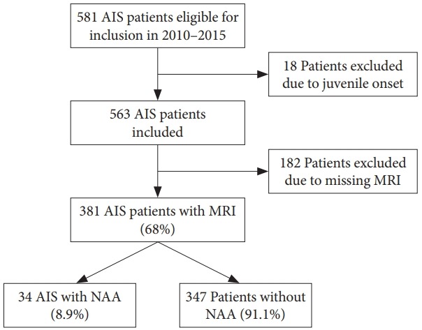
Methods: We prospectively included AIS patients referred to a tertiary facility for evaluation. Full-spine MRI scans were performed on all included patients irrespective of curve magnitude or proposed treatment modality. MRI scans were prospectively analyzed by a neuroradiologist and the pathologic findings were confirmed by a second independent radiologist.
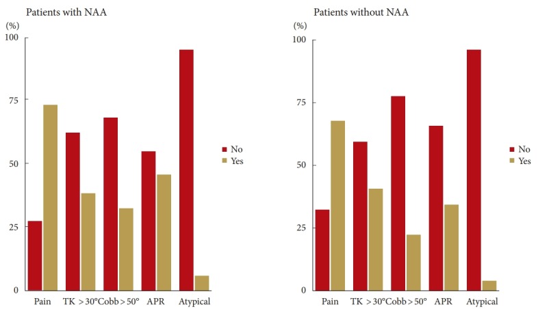
Results: NAA was observed in 34 of the 381 patients (8.9%): 32 patients had a syrinx, 1 patient had an arachnoid cyst, and 1 patient had a Chiari malformation. Four patients were referred for a neurosurgical evaluation but none received any neurosurgical treatment. No statistically significant difference was observed between the NAA and non-NAA groups in terms of sex, major curve size, thoracic kyphosis, left thoracic curve, curve convexity, curve progression, or level of pain (p>0.05).
Conclusion: In this prospective study examining the risk factors for NAA in AIS patients, we found that previously proposed risk factors could not predict the MRI outcomes. The finding of an NAA had no clinical implications and we do not support MRI scans as a routine diagnostic modality in all AIS patients.
Keywords: Adolescent idiopathic scoliosis; Magnetic resonance imaging; Neural axis abnormality; Syringomyelia; Syrinx.
Conflict of interest statement
Benny Dahl received institutional grants from K2M and MEDTRONIC outside the submitted work. Martin Gehrchen received institutional grants from K2M and MEDTRONIC outside the submitted work. Sidsel Fruergaard received institutional grants from MEDTRONIC. Søren Ohrt-Nissen received institutional grants from K2M outside the submitted work. The other authors have nothing to disclose.
Figures

References
-
- Do T, Fras C, Burke S, et al. Clinical value of routine preoperative magnetic resonance imaging in adolescent idiopathic scoliosis. A prospective study of three hundred and twenty-seven patients. J Bone Joint Surg Am. 2001;83-A:577–9. - PubMed
-
- Benli IT, Uzümcügil O, Aydin E, et al. Magnetic resonance imaging abnormalities of neural axis in Lenke type 1 idiopathic scoliosis. Spine (Phila Pa 1976) 2006;31:1828–33. - PubMed
-
- Inoue M, Minami S, Nakata Y, et al. Preoperative MRI analysis of patients with idiopathic scoliosis: a prospective study. Spine (Phila Pa 1976) 2005;30:108–14. - PubMed
-
- Nakahara D, Yonezawa I, Kobanawa K, et al. Magnetic resonance imaging evaluation of patients with idiopathic scoliosis: a prospective study of four hundred seventy-two outpatients. Spine (Phila Pa 1976) 2011;36:E482–5. - PubMed
LinkOut - more resources
Full Text Sources
