

Lumbar Intramedullary Epidermoid Following Repair of Sacral Myelomeningocele and Tethered Cord: A Case Report With a Review of the Relevant Literature and Operative Nuances
- PMID: 30653913
- PMCID: PMC6603839
- DOI: 10.14245/ns.1836152.076
Lumbar Intramedullary Epidermoid Following Repair of Sacral Myelomeningocele and Tethered Cord: A Case Report With a Review of the Relevant Literature and Operative Nuances
Abstract
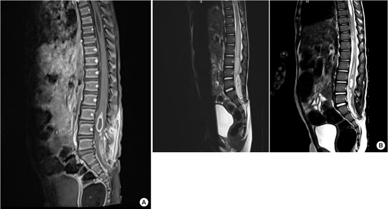
Epidermoid cysts of the spine are rare tumors. While the majority of them occur spontaneously, in very few cases, they can occur following previous surgery for spinal dysraphism. Such tumors tend to occur at the site of previous surgery. The occurrence of an epidermoid cyst at a level higher than the previous surgical site is a rare entity. We present a rare case of a lumbar intramedullary and extramedullary epidermoid occurring at a level higher than the previous surgical site, along with a discussion of the causes of such an occurrence and operative nuances regarding the management of an intramedullary epidermoid in a pediatric patient.
Keywords: Epidermoid cyst; Intramedullary epidermoid; Spinal dysraphism.
Conflict of interest statement
The authors have nothing to disclose.
Figures
Similar articles
-
Dorsal Spinal Intradural Intramedullary Epidermoid Cyst: A Rare Case Report and Review of Literature.J Neurosci Rural Pract. 2019 Apr-Jun;10(2):352-354. doi: 10.4103/jnrp.jnrp_304_18. J Neurosci Rural Pract. 2019. PMID: 31001035 Free PMC article.
-
Surgical Resection of a Recurrent Intramedullary Spinal Epidermoid Cyst: 2-Dimensional Operative Video.Oper Neurosurg. 2019 Nov 1;17(5):E204. doi: 10.1093/ons/opz019. Oper Neurosurg. 2019. PMID: 30851048
-
Dorsal intramedullary spinal epidermoid cysts: Report of two cases and review of literature.Indian J Orthop. 2007 Oct;41(4):395-7. doi: 10.4103/0019-5413.37005. Indian J Orthop. 2007. PMID: 21139798 Free PMC article.
-
A rare case of intradural and extramedullary epidermoid cyst after repetitive epidural anesthesia: case report and review of the literature.World J Surg Oncol. 2017 Jul 17;15(1):131. doi: 10.1186/s12957-017-1186-4. World J Surg Oncol. 2017. PMID: 28716031 Free PMC article. Review.
-
Spinal Intramedullary Epidermoid Cyst: Case Report and Updated Literature Review.World Neurosurg. 2020 Jul;139:39-50. doi: 10.1016/j.wneu.2020.03.207. Epub 2020 Apr 13. World Neurosurg. 2020. PMID: 32298825 Review.
Cited by
-
Ossified spinal epidermoid cyst: A systematic review and case report.Heliyon. 2024 Aug 28;10(18):e37093. doi: 10.1016/j.heliyon.2024.e37093. eCollection 2024 Sep 30. Heliyon. 2024. PMID: 39315203 Free PMC article.
-
Surgical Strategy for Sacral Tumor Resection.Yonsei Med J. 2021 Jan;62(1):59-67. doi: 10.3349/ymj.2021.62.1.59. Yonsei Med J. 2021. PMID: 33381935 Free PMC article.
References
-
- Mohapatra RN, Patra RK, Panigrahi MK. Cauda equina syndrome due to implantation epidermoid tumor: after 38 years of surgery for lumbar meningocele in a neonate. J Pediatr Neurosci. 2007;2:28–30.
-
- Ogden AT, Khandji AG, McCormick PC, et al. Intramedullary inclusion cysts of the cervicothoracic junction. Report of two cases in adults and review of the literature. J Neurosurg Spine. 2007;7:236–42. - PubMed
-
- Park JC, Chung CK, Kim HJ. Iatrogenic spinal epidermoid tumor. A complication of spinal puncture in an adult. Clin Neurol Neurosurg. 2003;105:281–5. - PubMed
-
- Kachhara R, Unnikrishnan M. Acquired cauda equina epidermoid cyst. Neurol India. 1999;47:250. - PubMed
Publication types
LinkOut - more resources
Full Text Sources
