. 2019 Jan 16;8(1):24.
doi: 10.3390/antiox8010024.

Effects of N-Acetyl-Cysteine Supplementation through Drinking Water on the Glutathione Redox Status during the Weaning Transition of Piglets

Affiliations

Effects of N-Acetyl-Cysteine Supplementation through Drinking Water on the Glutathione Redox Status during the Weaning Transition of Piglets

Jeroen Degroote et al. Antioxidants (Basel). .

Abstract

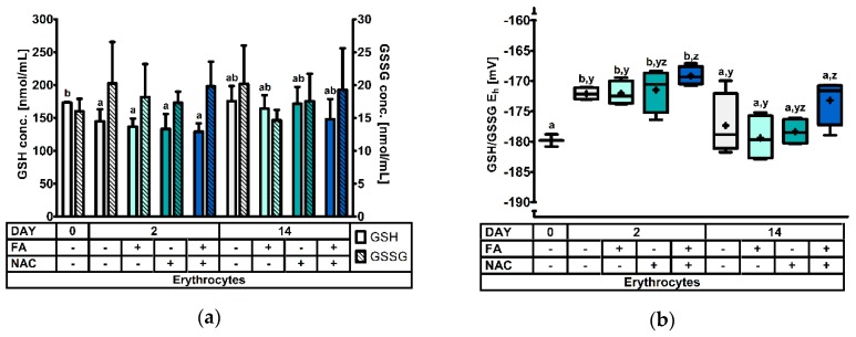
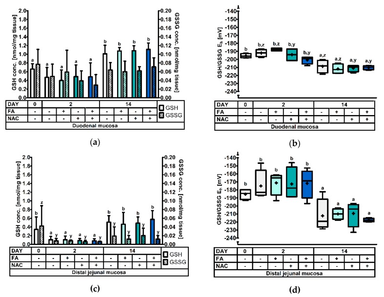
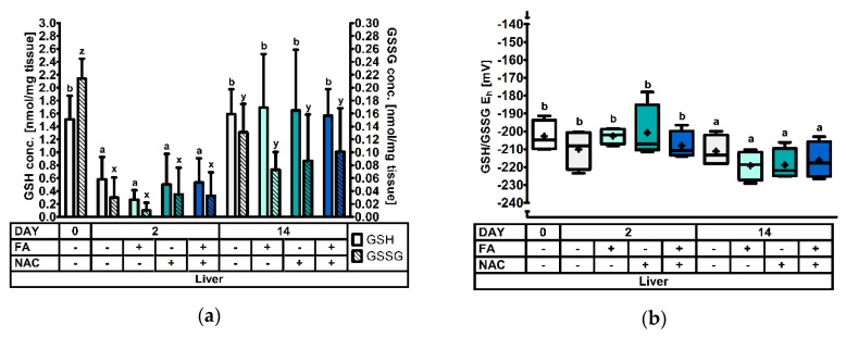
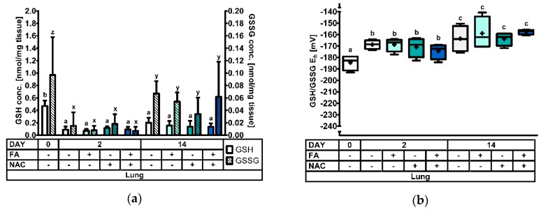
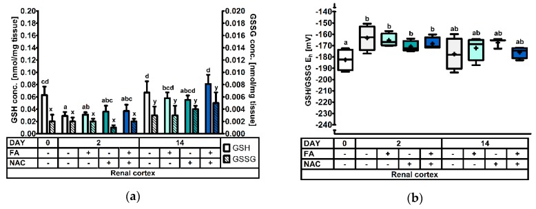
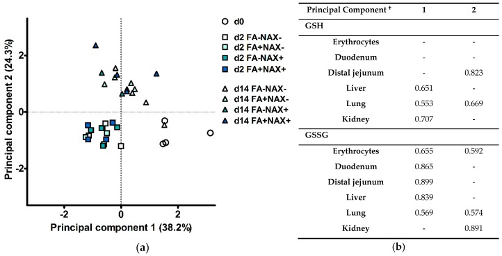
This study investigated the effect of N-acetyl-cysteine (NAC) supplementation through drinking water on animal performance and the glutathione (GSH) redox system in weaned piglets, particularly in relation to the immediate post-weaning feed intake. To this end, 168 piglets were weaned and either fed ad libitum or fasted the first two days, and either or not administered 200 mg/L NAC via the drinking water until d14 post-weaning. Next to animal performance until day 42 (d42), the GSH redox system was measured in erythrocytes, small intestinal mucosa, liver, lung, and kidney tissue at d0, d2, and d14 post-weaning. Animal performance and GSH levels were not affected by NAC, nor by fasting. Irrespective of treatment, a significant drop in GSH at d2 post-weaning was found as compared to d0, in particular in liver (-69%), distal jejunal mucosa (-72%), and lung tissue (-80%). Post-weaning changes of the GSH redox status were strongly tissue-dependent. To conclude, this research indicates that GSH redox homeostasis was largely affected in multiple organs during the weaning transition. NAC supplementation did not increase GSH levels in any tissue, not even in fasted animals, questioning the fact if cysteine is the first or only limiting factor determining the rate of GSH synthesis in the early post-weaning phase.

Keywords: N-acetyl-cysteine; glutathione; redox status; small intestine; weaned pigs.


Conflict of interest statement

The authors declare no conflict of interest. The funders had no role in the design of the study; in the collection, analyses, or interpretation of data; in the writing of the manuscript, or in the decision to publish the results.

Figures

Figure 1
Figure 1
The glutathione redox system in erythrocytes of piglets at d0, d2, and d14 post-weaning (DAY), either ad libitum fed (FA−) or fasted (FA+) the first 2d post-weaning, and either provided standard drinking water (NAC−) or water supplemented with 200 mg/L N-acetyl cysteine (NAC+) during the first 14d post-weaning. Results are presented as least squares means with SD. Significance levels of main effects and interaction terms are presented in Table 3: (a) Erythrocyte glutathione (GSH) and glutathione disulphide (GSSG) concentrations. a,b Represent the effect of DAY across other factors (p ≤ 0.05); (b) Erythrocyte glutathione redox status (GSH/GSSG Eh). a,b Represent the effect of DAY across other factors (p ≤ 0.05). y,z Represent the effect of FA × NAC across DAYS (p ≤ 0.05).
Figure 2
Figure 2
The glutathione redox system in the small intestinal mucosa of piglets at 0d, 2d, and 14d post-weaning (DAY), either ad libitum fed (FA−) or fasted (FA+) the first 2d post-weaning, and either provided standard drinking water (NAC−) or water supplemented with 200 mg/L N-acetyl cysteine (NAC+) during the first 14d post-weaning. Results are presented as least squares means with SD. Significance levels of main effects and interaction terms are presented in Table 3: (a,c) Glutathione (GSH) and glutathione disulphide (GSSG) concentrations in the duodenal mucosa (a) or the distal jejunal mucosa (c). a,b And y,z respectively represent the effect of DAY on GSH and GSSG concentrations across other factors (p ≤ 0.05); (b,d) Glutathione redox status (GSH/GSSG Eh) in the duodenal mucosa (b) or the distal jejunal mucosa (d). a,b and y,z respectively represent the effect of DAY and NAC across other factors (p ≤ 0.05).
Figure 3
Figure 3
The glutathione redox system in liver tissue of piglets at 0d, 2d, and 14d post-weaning (DAY), either ad libitum fed (FA−) or fasted (FA+) the first 2d post-weaning, and either provided standard drinking water (NAC−) or water supplemented with 200 mg/L N-acetyl cysteine (NAC+) during the first 14d post-weaning. Results are presented as least squares means with SD. Significance levels of main effects and interaction terms are presented in Table 3: (a) Hepatic glutathione (GSH) and glutathione disulphide (GSSG) concentrations. a,b and x,y,z respectively represent the effect of DAY on GSH or GSSG concentrations across other factors (p ≤ 0.05); (b) Hepatic glutathione redox status (GSH/GSSG Eh). a,b represent the effect of DAY across other factors (p ≤ 0.05).
Figure 4
Figure 4
The glutathione redox system in lung tissue of piglets at 0d, 2d, and 14d post-weaning (DAY), either ad libitum fed (FA−) or fasted (FA+) the first 2d post-weaning, and either provided standard drinking water (NAC−) or water supplemented with 200 mg/L N-acetyl cysteine (NAC+) during the first 14d post-weaning. Results are presented as least squares means with SD. Significance levels of main effects and interaction terms are presented in Table 3: (a) Pulmonary glutathione (GSH) and glutathione disulphide (GSSG) concentrations. a,b and x,y,z respectively represent the effect of DAY on GSH or GSSG concentrations across other factors (p ≤ 0.05); (b) Pulmonary glutathione redox status (GSH/GSSG Eh). a,b,c represent the effect of DAY across other factors (p ≤ 0.05).
Figure 5
Figure 5
The glutathione redox system in kidney tissue of piglets at 0d, 2d and 14d post-weaning (DAY), either ad libitum fed (FA−) or fasted (FA+) the first 2d post-weaning, and either provided standard drinking water (NAC−) or water supplemented with 200 mg/L N-acetyl cysteine (NAC+) during the first 14d post-weaning. Results are presented as least squares means with SD. Significance levels of main effects and interaction terms are presented in Table 3: (a) Renal cortical glutathione (GSH) and glutathione disulphide (GSSG) concentrations. a,b,c,d and x,y respectively represent differences in GSH or GSSG concentrations between treatments (p ≤ 0.05); (b) Renal cortical glutathione redox status (GSH/GSSG Eh). a,b represent the effect of DAY across other factors (p ≤ 0.05).
Figure 6
Figure 6
Principal component analysis of glutathione (GSH) and glutathione disulfide (GSSG) concentrations in six different tissues of piglets at 0d, 2d, and 14d post-weaning, either ad libitum fed (FA−) or fasted (FA+) the first 2d post-weaning, and either provided standard drinking water (NAC−) or water supplemented with 200 mg/L N-acetyl cysteine (NAC+) during the first 14d post-weaning: (a) Scores plot representing the 36 individual piglets in the multivariate space of the first two principal components. Animals can be visually clustered according to day post-weaning (d0, d2, or d14). (b) Loadings of the two major principal components. These loadings indicate the correlation (positive or negative) between the original variables and the principal component. Squared factor loadings indicate the percentage of variance in an original variable explained by the principal component. Only correlations with |r|> 0.5 are indicated.

Similar articles

Cited by

References

    1. Degroote J., Michiels J., Claeys E., Ovyn A., De Smet S. Changes in the pig small intestinal mucosal glutathione kinetics after weaning. J. Anim. Sci. 2012;90:359–361. doi: 10.2527/jas.53809. - DOI - PubMed
    1. Luo Z., Zhu W., Guo Q., Luo W., Zhang J., Xu W., Xu J. Weaning Induced Hepatic Oxidative Stress, Apoptosis, and Aminotransferases through MAPK Signaling Pathways in Piglets. Oxidative Med. Cell. Longev. 2016;2016:1–10. doi: 10.1155/2016/4768541. - DOI - PMC - PubMed
    1. Jones D.P., Sies H. The Redox Code. Antioxid. Redox Signal. 2015;23:734–746. doi: 10.1089/ars.2015.6247. - DOI - PMC - PubMed
    1. Wu G., Fang Y.Z., Yang S., Lupton J.R., Turner N.D. Glutathione metabolism and its implications for health. J. Nutr. 2004;134:489–492. doi: 10.1093/jn/134.3.489. - DOI - PubMed
    1. Circu M.L., Aw T.Y. Intestinal redox biology and oxidative stress. Semin. Cell Dev. Biol. 2012;23:729–737. doi: 10.1016/j.semcdb.2012.03.014. - DOI - PMC - PubMed

LinkOut - more resources