. 2019 Jan 16;20(2):345.
doi: 10.3390/ijms20020345.

Cellular and Molecular Mechanisms Mediated by recPrPC Involved in the Neuronal Differentiation Process of Mesenchymal Stem Cells

Affiliations

Cellular and Molecular Mechanisms Mediated by recPrPC Involved in the Neuronal Differentiation Process of Mesenchymal Stem Cells

Stefano Martellucci et al. Int J Mol Sci. .

Abstract

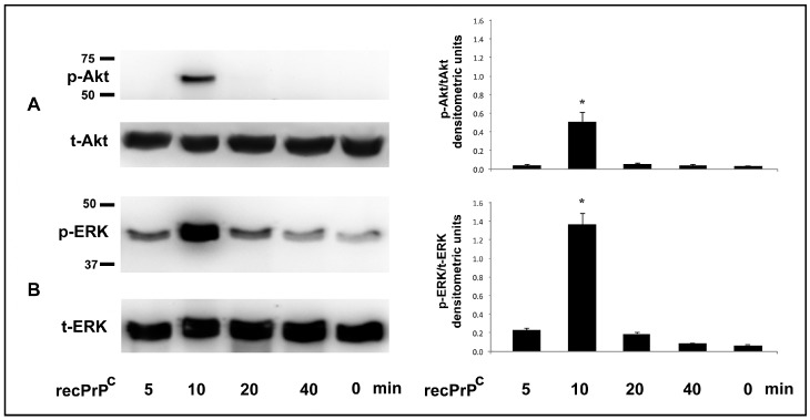
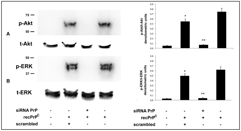
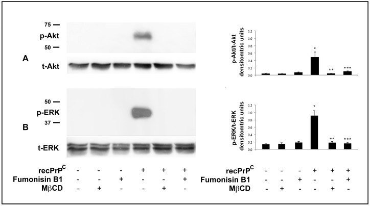
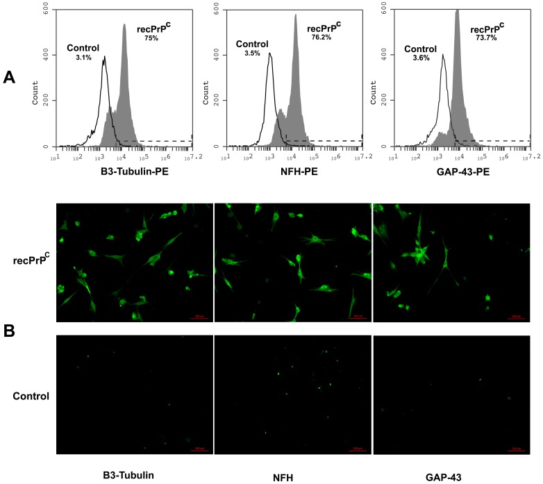
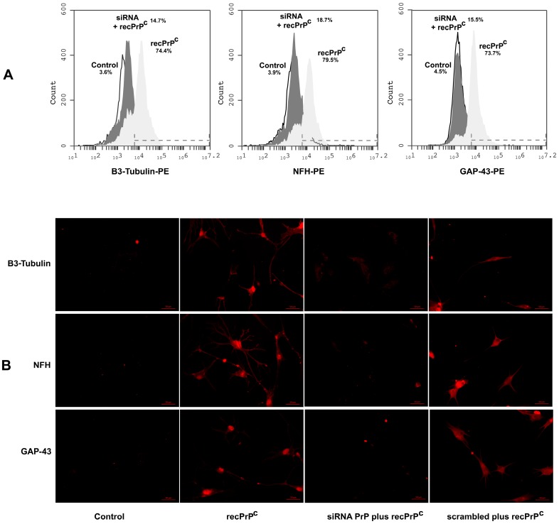
Human Dental Pulp Stem Cells (hDPSCs) represent a type of adult mesenchymal stem cells that have the ability to differentiate in vitro in several lineages such as odontoblasts, osteoblasts, chondrocytes, adipocytes and neurons. In the current work, we used hDPSCs as the experimental model to study the role of recombinant prion protein 23⁻231 (recPrPC) in the neuronal differentiation process, and in the signal pathway activation of ERK 1/2 and Akt. We demonstrated that recPrPC was able to activate an intracellular signal pathway mediated by extracellular-signal-regulated kinase 1 and 2 (ERK 1/2) and protein kinase B (Akt). Moreover, in order to understand whether endogenous prion protein (PrPC) was necessary to mediate the signaling induced by recPrPC, we silenced PrPC, demonstrating that the presence of endogenous PrPC was essential for ERK 1/2 and Akt phosphorylation. Since endogenous PrPC is a well-known lipid rafts component, we evaluated the role of these structures in the signal pathway induced by recPrPC. Our results suggest that lipid rafts integrity play a key role in recPrPC activity. In fact, lipid rafts inhibitors, such as fumonisin B1 and MβCD, significantly prevented ERK 1/2 and Akt phosphorylation induced by recPrPC. In addition, we investigated the capacity of recPrPC to induce hDPSCs neuronal differentiation process after long-term stimulation through the evaluation of typical neuronal markers expression such as B3-Tubulin, neurofilament-H (NFH) and growth associated protein 43 (GAP43). Accordingly, when we silenced endogenous PrPC, we observed the inhibition of neuronal differentiation induced by recPrPC. The combined data suggest that recPrPC plays a key role in the neuronal differentiation process and in the activation of specific intracellular signal pathways in hDPSCs.

Keywords: adult neurogenesis; cellular prion protein; dental pulp-derived stem cells; lipid rafts; mesenchymal stem cells; neural stem cells; neuronal differentiation; prions; recombinant prion protein; shed prion protein.


Conflict of interest statement

The authors declare no conflicts of interest.

Figures

Figure 1
Figure 1
Effect of recPrPC on Akt and ERK Phosphorylation. hDPSCs, untreated or treated with 0.5 µg/mL of recPrPC for 5, 10, 20, 40 min, were analyzed by Western blot using anti-pAkt, anti-total Akt (A). anti-pERK1/2 and anti-total ERK1/2 (B). Densitometric analysis is shown in the right panel. Results represent the mean ± SD from 3 independent experiments, * p recPrPC treated cells <0.01 vs. untreated cells.
Figure 2
Figure 2
Effect of PrPC silencing on Akt and ERK phosphorylation induced by recPrPC. hDPSCs, untreated or treated with 0.5 µg/mL of recPrPC for 10 min in presence or in absence of pre-treatment with siRNA PrP or scrambled siRNA for 72 h, were analyzed by Western blot using anti-pAkt, anti-total Akt (A). anti-pERK1/2 and anti-total ERK1/2 (B). Densitometric analysis is shown in the right panel. Results represent the mean ± SD from 3 independent experiments, * p recPrPC treated cells <0.01 vs. untreated cells, ** p siRNA PrP + recPrPC treated cells vs. scrambled + recPrPC treated cells. As control, scrambled siRNA was employed in each experiment.
Figure 3
Figure 3
Effect of lipid rafts perturbation on Akt and ERK Phosphorylation induced by recPrPC. hDPSCs, untreated or treated with 0.5 µg/mL of recPrPC for 10 min in the presence or in the absence of Fumonisin B1 or MβCD, were analyzed by Western blot using anti-pAkt and anti-total Akt (A). anti-pERK1/2 and anti-total ERK1/2 (B). Densitometric analysis is shown in the right panel. Results represent the mean ± SD from 3 independent experiments, * p recPrPC treated cells <0.01 vs. untreated cells, ** p recPrPC treated cells + MβCD <0.01 vs. recPrPC treated cells, *** p recPrPC treated cells + Fumonisin B1 <0.01 vs. recPrPC treated cells.
Figure 4
Figure 4
Effect of recPrPC on expression of neuronal markers GAP-43, NFH and B3-Tubulin. hDPSCs, untreated or treated with recPrPC for 14 days, were analyzed by flow cytometry and immunofluorescence analysis using anti-NFH, Anti-B3-Tubulin and anti-GAP-43. (A) Flow Cytometry. Histograms represent log fluorescence vs. cell number, gated on cell population of a side scatter/forward scatter (SS/FS) histogram. Cell number is indicated on the y-axis and fluorescence intensity is represented on the x-axis. Each panel was compared with the corresponding secondary antibody as negative control. A representative experiment among 3 is shown. (B) Immunofluorescence analysis. Alternatively, hDPSCs were analyzed by immunofluorescence analysis using anti-B3-Tubulin, anti-NFH and anti-GAP-43 and observed with a Zeiss Axio Vert. A1 fluorescence microscope (Zeiss, Oberkochen, Germany). Scale bars, 100 µm.
Figure 5
Figure 5
Effect of PrPC silencing on expression of neuronal markers B3-Tubulin, NFH and GAP-43. hDPSCs, stimulated with 0.5 µg/mL of recPrPC for 14 days in the presence or in the absence of pre-treatment with siRNA PrP or scrambled siRNA for 72 h, were analyzed by flow cytometry or immunofluorescence analysis using anti-B3-Tubulin, anti-NFH and anti-GAP-43. (A) Flow Cytometry. Histograms represent log fluorescence vs. cell number, gated on cell population of a side scatter/forward scatter (SS/FS) histogram. Cell number is indicated on the y-axis, and fluorescence intensity is represented on the x-axis. Each panel was compared with the corresponding secondary antibody as a negative control. A representative experiment among 3 is shown. (B) Immunofluorescence analysis. Alternatively, hDPSCs were analyzed by immunofluorescence analysis using anti-B3-Tubulin, anti-NFH and anti-GAP-43 and, then, observed with a Zeiss Axio Vert. A1 fluorescence microscope. Scale bars, 50 µm.

Similar articles

Cited by

References

    1. Brown D.R., Qin K., Herms J.W., Madlung A., Manson J., Strome R., Fraser P.E., Kruck T., von Bohlen A., Schulz-Schaeffer W., et al. The cellular prion protein binds copper in vivo. Nature. 1997;390:684–687. doi: 10.1038/37783. - DOI - PubMed
    1. Mattei V., Martellucci S., Santilli F., Manganelli V., Garofalo T., Candelise N., Caruso A., Sorice M., Scaccianoce S., Misasi R. Morphine Withdrawal Modifies Prion Protein Expression in Rat Hippocampus. PLoS ONE. 2017;12:e0169571. doi: 10.1371/journal.pone.0169571. - DOI - PMC - PubMed
    1. Lewis V., Hooper N.M. The role of lipid rafts in prion protein biology. Front Biosci. 2011;16:151–168. doi: 10.2741/3681. - DOI - PubMed
    1. Sorice M., Mattei V., Tasciotti V., Manganelli V., Garofalo T., Misasi R. Trafficking of PrPC to mitochondrial raft-like microdomains during cell apoptosis. Prion. 2012;6:354–358. doi: 10.4161/pri.20479. - DOI - PMC - PubMed
    1. Prusiner S.B., Scott M.R., DeArmond S.J., Cohen F.E. Prion protein biology. Cell. 1998;93:337–348. doi: 10.1016/S0092-8674(00)81163-0. - DOI - PubMed

MeSH terms