. 2019 Jan 16;20(2):363.
doi: 10.3390/ijms20020363.

AZD1208, a Pan-Pim Kinase Inhibitor, Has Anti-Growth Effect on 93T449 Human Liposarcoma Cells via Control of the Expression and Phosphorylation of Pim-3, mTOR, 4EBP-1, S6, STAT-3 and AMPK

Affiliations

AZD1208, a Pan-Pim Kinase Inhibitor, Has Anti-Growth Effect on 93T449 Human Liposarcoma Cells via Control of the Expression and Phosphorylation of Pim-3, mTOR, 4EBP-1, S6, STAT-3 and AMPK

Anil Kumar Yadav et al. Int J Mol Sci. .

Abstract

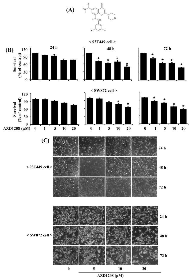
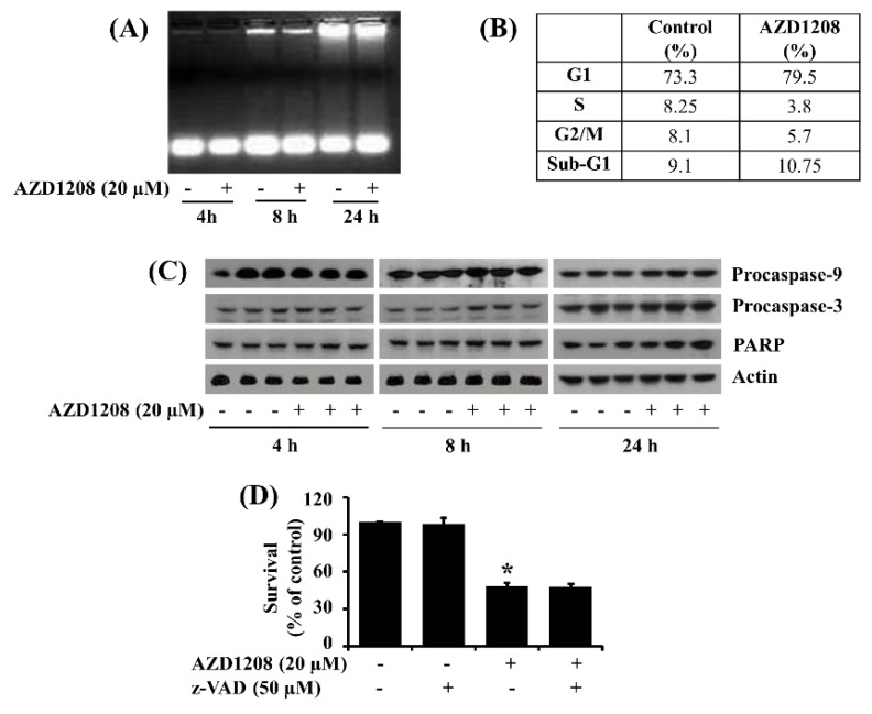
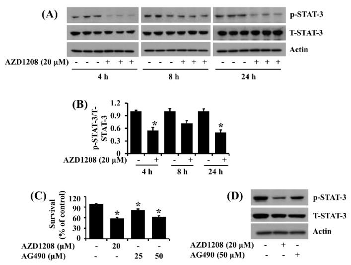
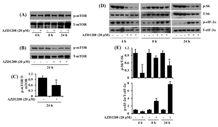
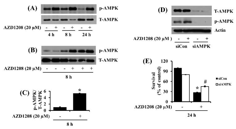
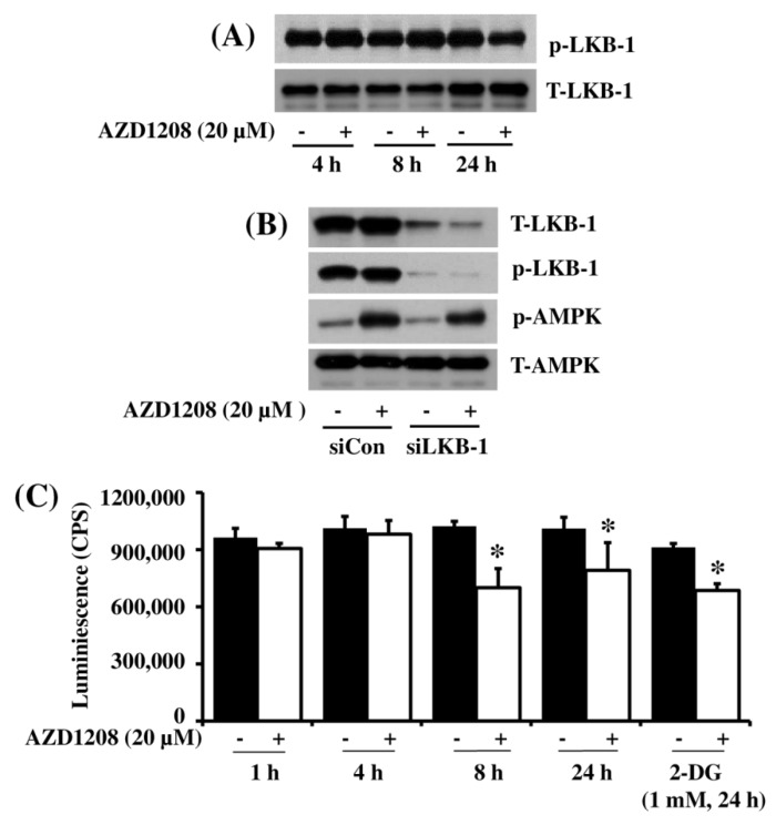
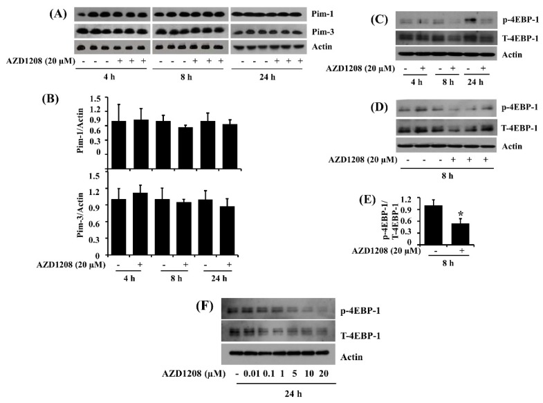
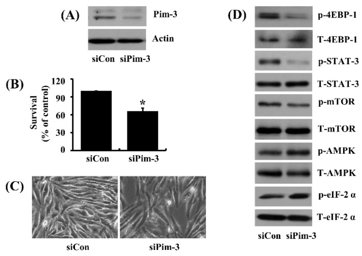
Overexpression of Pim kinases has an oncogenic/pro-survival role in many hematological and solid cancers. AZD1208 is a pan-Pim kinase inhibitor that has anti-cancer and anti-adipogenic actions. Here, we investigated the effects of AZD1208 on the growth of 93T449 cells, a differentiated human liposarcoma cell line. At 20 µM, AZD1208 was cytotoxic (cytostatic) but not apoptotic, reducing cell survival without DNA fragmentation, caspase activation or increasing cells in the sub G1 phase; known apoptotic parameters. Notably, AZD1208 reduced phosphorylation of signal transducer and activator of transcription-3 (STAT-3) in 93T449 cells. STAT-3 inhibition by AG490, a JAK2/STAT-3 inhibitor similarly reduced cell survival. AZD1208 down-regulated phosphorylation of mammalian target of rapamycin (mTOR) and ribosomal S6 while up-regulated eukaryotic initiation factor-2α (eIF-2α). In addition, AZD1208 induced a LKB-1-independent AMPK activation, which was crucial for its cytostatic effect, as knock-down of AMPK greatly blocked AZD1208s ability to reduce cell survival. AZD1208 had no effect on expression of two members of Pim kinase family (Pim-1 and Pim-3) but inhibited phosphorylation of 4EBP-1, a downstream effector of Pim kinases. Importantly, a central role for Pim-3 in the actions of AZD1208 was confirmed by knock-down, which not only reduced 93T449 cell survival but also led to the inhibition of 4EBP-1, mTOR, eIF-2α and STAT-3, along with the activation of AMPK. In summary, this is the first report demonstrating that AZD1208 inhibits growth of liposarcoma cells and that this activity is mediated through Pim-3 kinase, STAT-3, mTOR, S6 and AMPK expression and phosphorylation pathways.

Keywords: 93T449; AMPK; AZD1208; Pim-3; STAT-3.


Conflict of interest statement

The authors declare no conflict of interest.

Figures

Figure 1
Figure 1
Effect of AZD1208 on survival of 93T449 and SW872 cells. (A) The chemical structure of AZD1208. (B) 93T449 and SW872 cells were treated with AZD1208 or vehicle control (DMSO; 0.1%) at the indicated concentrations and for the indicated times. The numbers of surviving cells were measured by cell count assay. The cell count assay was performed in triplicate. Data are means ± SE of three independent experiments. * p < 0.05 compared to the value of AZD1208 free control at the indicated time. (C) 93T449 and SW872 cells were treated with AZD1208 or vehicle control (DMSO) for the indicated times. Images of the conditioned cells were obtained by phase contrast microscopy, 200 ×. Each image is a representative of three independent experiments.
Figure 2
Figure 2
Effect of AZD1208 on apoptosis of 93T449 cells. (A) 93T449 cells were treated with AZD1208 (20 µM) or vehicle control (DMSO) for the times indicated. At each time point, extra-nuclear fragmented DNA from the conditioned cells was extracted and analyzed on a 1.7% agarose gel. The image is a representative of three independent experiments. (B) 93T449 cells were treated with AZD1208 (20 µM) or vehicle control (DMSO) for 24 h. The conditioned cells were harvested and subjected to fluorescence-activated cell sorting (FACS) analysis for measuring the population of sub G1 phase. The tables represent the fraction of apoptotic cells. (C) 93T449 cells were treated with AZD1208 (20 µM) or vehicle control (DMSO) in triplicate experiments for the times designated. At each time point, whole cell lysates were prepared and analyzed for procaspase-9, procaspase-3, PARP or β-actin expression or cleavage by Western blotting. (D) 93T449 cells were treated without or with AZD1208 (20 µM) in the absence or presence of the pan-caspase inhibitor z-VAD (50 µM) for 48 h, followed measurement of the number of surviving cells by cell count assay. The cell count assay was done in triplicate. Data are means ± SE of three independent experiments. * p < 0.05 compared to the control at the indicated time.
Figure 3
Figure 3
Effect of AZD1208 on expression and phosphorylation levels of STAT-3 in 93T449 cells. (A) 93T449 cells were treated with AZD1208 (20 µM) or vehicle control (DMSO) in triplicate experiments for the times designated. At each time point, whole cell lysates were prepared and analyzed for levels of p-STAT-3, T-STAT-3 or β-actin by Western blotting. p-STAT-3, phosphorylated STAT-3; T-STAT-3, total STAT-3. (B) The densitometry data of (A). * p < 0.05 compared to the control at the indicated time. (C) 93T449 cells were treated with AZD1208 or AG490, a Jak/STAT-3 inhibitor, at the indicated concentrations for 48 h, followed measurement of the number of surviving cells by cell count assay. The cell count assay was performed in triplicate. Data are means ± SE of three independent experiments. (D) 93T449 cells were treated with AZD1208 (20 µM) or AG490 (50 µM) for 24 h. Whole cell lysates from the conditioned cells were prepared and analyzed for p-STAT-3, T-STAT-3 or β-actin by Western blotting. The image is a representative of three independent experiments.
Figure 4
Figure 4
Effect of AZD1208 on expression and phosphorylation of mTOR, S6 and eIF-2α in 93T449 cells. (A) 93T449 cells were treated with AZD1208 (20 µM) or vehicle control (DMSO) for the times designated. At each time point, whole cell lysates were prepared and analyzed for p-mTOR and T-mTOR by Western blotting. p-mTOR, phosphorylated mTOR; T-mTOR, total mTOR. (B) Western blotting analysis in triplicate experiments for 24 h. (C) The densitometry data of (B). * p < 0.05 compared to the control at the indicated time. (D) 93T449 cells were treated with AZD1208 (20 µM) or vehicle control (DMSO) in triplicate experiments for indicated times. At each time point, whole cell lysates were prepared and analyzed for p-S6, T-S6, p-eIF-2α and T-eIF-2α by Western blotting. p-S6, phosphorylated S6; T-S6, total S6; p-eIF-2α, phosphorylated eIF-2α; T-eIF-2α, total eIF-2α. (E) The densitometry data of (D). * p < 0.05 compared to the control at the indicated time.
Figure 5
Figure 5
Effect of AZD1208 and/or knock-down of AMPK on survival of 93T449 cells and cellular expression and phosphorylation of AMPK. (A) 93T449 cells were treated with AZD1208 (20 µM) or vehicle control (DMSO) for the times designated. At each time point, whole cell lysates were prepared and analyzed for p-AMPK and T-AMPK by Western blotting. p-AMPK, phosphorylated AMPK; T-AMPK, total AMPK. (B) Western blotting in triplicate experiments for 8 h. (C) The densitometry data of (B). * p < 0.05 compared to the control at the indicated time. (D) 93T449 cells were transfected with 100 pM of control siRNA (siCon) or AMPK siRNA (siAMPK) for 24 h. The siCon- or siAMPK-transfected cells were treated with AZD1208 (20 µM) or vehicle control for 24 h. Whole cell lysates were prepared and analyzed for p-AMPK, T-AMPK or β-actin by Western blotting. (E) 93T449 cells were transfected with 100 pM of control siRNA (siCon) or AMPK siRNA (siAMPK) for 24 h. The siCon- or siAMPK-transfected cells were treated with AZD1208 (20 µM) or vehicle control for 24 h, followed measurement of the number of surviving cells by cell count assay. The cell count assay was performed in triplicate. Data are means ± SE of three independent experiments. * p < 0.05 compared to the control at the indicated time; # p < 0.05 compared to AZD1208 at the indicated time.
Figure 6
Figure 6
Effect of AZD1208 and/or knock-down of LKB-1 on expression and phosphorylation of LKB-1 and AMPK and cellular ATP content in 93T449 cells. (A) 93T449 cells were treated with AZD1208 (20 µM) or vehicle control (DMSO) for the times designated. At each time point, whole cell lysates were prepared and analyzed for p-LKB-1 and T-LKB-1 by Western blotting. p-LKB-1, phosphorylated LKB-1; T-LKB-1, total LKB-1. The image is a representative of three independent experiments. (B) 93T449 cells were transfected with 100 pM of control siRNA (siCon) or LKB-1 siRNA (siLKB-1) for 24 h. The siCon- or siAMPK-transfected cells were treated with AZD1208 (20 µM) or vehicle control for 24 h. Whole cell lysates were prepared and analyzed for T-LKB-1, p-LKB-1, p-AMPK and T-AMPK by Western blotting. (C) 93T449 cells were treated with AZD1208 (20 µM) or vehicle control for the times designated. For comparison, cells were treated with 1 mM of deoxyglucose (2-DG), a known ATP depleting agent for 24 h. At each time point, cellular ATP content was measured by ATP measurement kit. * p < 0.05 compared to the value of AZD1208 or 2-DG free control at the indicated time.
Figure 7
Figure 7
Effect of AZD1208 on expression and/or phosphorylation of Pim kinases and 4EBP-1 in 93T449 cells. (A) 93T449 cells were treated with AZD1208 (20 µM) or vehicle control (DMSO) in triplicate experiments for the times designated. At each time point, whole cell lysates were prepared and analyzed for Pim-1, Pim-3 and β-actin by Western blotting. (B) The densitometry data of (A). (C) 93T449 cells were treated with AZD1208 (20 µM) or vehicle control for the times designated. At each time point, whole cell lysates from the conditioned cells were prepared and analyzed for p-4EBP-1, T-4EBP-1 and β-actin by Western blotting. p-4EBP-1, phosphorylated 4EBP-1; T-4EBP-1, total 4EBP-1. (D) Western blotting in triplicate experiments for 8 h. (E) The densitometry data of (D). * p < 0.05 compared to the value of AZD1208 free control at the indicated time (F) 93T449 cells were treated with different concentrations of AZD1208 (0.01, 0.1, 1, 5, 10 and 20 µM) or vehicle control for 24 h. Whole cell lysates from the conditioned cells were prepared and analyzed for p-4EBP-1, T-4EBP-1 and β-actin by Western blotting. p-4EBP-1, phosphorylated 4EBP-1; T-4EBP-1, total 4EBP-1.
Figure 8
Figure 8
Effect of Pim-3 knock-down on survival of 93T449 cells and cellular expression and/or phosphorylation of 4EBP-1, STAT-3, AMPK, mTOR and eIF-2α. (A) 93T449 cells were transfected with 100 pM of control siRNA (siCon) or Pim-3 siRNA (siPim-3) for 24 h. Whole cell lysates were prepared and analyzed for Pim-3 and β-actin by Western blotting. (B) 93T449 cells were transfected with 100 pM of control siRNA (siCon) or Pim-3 siRNA (siPim-3) for 24 h, followed measurement of the number of surviving cells by cell count assay. The cell count assay was performed in triplicate. Data are means ± SE of three independent experiments. * p < 0.05 compared to the siConc at the indicated time. (C) Images of the siCon- or siPim-3-transfected cells were obtained by phase contrast microscopy, 400 ×. (D) 93T449 cells were transfected with 100 pM of control siRNA (siCon) or Pim-3 siRNA (siPim-3) for 24 h. Whole cell lysates were prepared and analyzed for p-4EBP-1, T-4EBP-1, p-STAT-3, T-STAT-3, p-AMPK, T-AMPK, p-mTOR, T-mTOR, p-eIF-2α and T-eIF-2α by Western blotting.

Similar articles

Cited by

References

    1. Cormier J.N., Pollock R.E. Soft tissue sarcomas, CA. Cancer J. Clin. 2004;54:94–109. doi: 10.3322/canjclin.54.2.94. - DOI - PubMed
    1. Dalal K.M., Antonescu C.R., Singer S. Diagnosis and Management of lipomatous tumors. J. Surg. Oncol. 2008;97:298–313. doi: 10.1002/jso.20975. - DOI - PubMed
    1. N.C. Institute Site, General Information About Adult Soft Tissue Sarcoma. [(accessed on 14 Januray 2018)]; Available online: https://www.cancer.gov/types/soft-tissue-sarcoma/hp/adult-soft-tissue-tr....
    1. Eilber F.C., Eilber F.R., Eckardt J., Rosen G., Riedel E., Maki R.G., Brennan M.F., Singer S. The impact of chemotherapy on the survival of patients with high-grade primary extremity liposarcoma. Ann. Surg. 2004;240:686–695. doi: 10.1097/01.sla.0000141710.74073.0d. - DOI - PMC - PubMed
    1. Singer S., Antonescu C.R., Riedel E., Brennan M.F. Histologic subtype and margin of resection predict pattern of recurrence and survival for retroperitoneal liposarcoma. Ann. Surg. 2003;238:358–370. doi: 10.1097/01.sla.0000086542.11899.38. - DOI - PMC - PubMed

MeSH terms

LinkOut - more resources