

Deep Learning Algorithms with Demographic Information Help to Detect Tuberculosis in Chest Radiographs in Annual Workers' Health Examination Data
- PMID: 30654560
- PMCID: PMC6352082
- DOI: 10.3390/ijerph16020250
Deep Learning Algorithms with Demographic Information Help to Detect Tuberculosis in Chest Radiographs in Annual Workers' Health Examination Data
Abstract
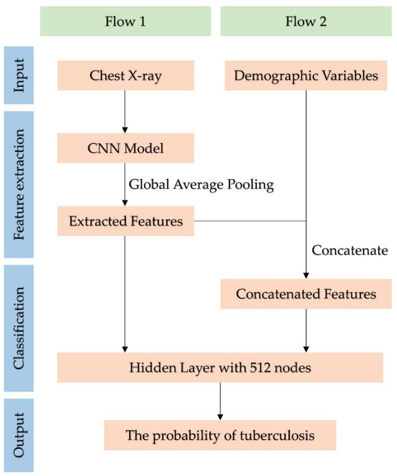
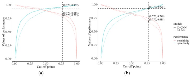
We aimed to use deep learning to detect tuberculosis in chest radiographs in annual workers' health examination data and compare the performances of convolutional neural networks (CNNs) based on images only (I-CNN) and CNNs including demographic variables (D-CNN). The I-CNN and D-CNN models were trained on 1000 chest X-ray images, both positive and negative, for tuberculosis. Feature extraction was conducted using VGG19, InceptionV3, ResNet50, DenseNet121, and InceptionResNetV2. Age, weight, height, and gender were recorded as demographic variables. The area under the receiver operating characteristic (ROC) curve (AUC) was calculated for model comparison. The AUC values of the D-CNN models were greater than that of I-CNN. The AUC values for VGG19 increased by 0.0144 (0.957 to 0.9714) in the training set, and by 0.0138 (0.9075 to 0.9213) in the test set (both p < 0.05). The D-CNN models show greater sensitivity than I-CNN models (0.815 vs. 0.775, respectively) at the same cut-off point for the same specificity of 0.962. The sensitivity of D-CNN does not attenuate as much as that of I-CNN, even when specificity is increased by cut-off points. Conclusion: Our results indicate that machine learning can facilitate the detection of tuberculosis in chest X-rays, and demographic factors can improve this process.
Keywords: computer-assisted diagnosis; convolutional neural network; deep learning; image; tuberculosis.
Conflict of interest statement
The authors declare no conflict of interest. The funders had no role in the design of the study; in the collection, analyses, or interpretation of data; in the writing of the manuscript; or in the decision to publish the results.
Figures
References
-
- Song H., Nguyen A.-D., Gong M., Lee S. A review of computer vision methods for purpose on computer-aided diagnosis. J. Int. Soc. Simul. Surg. 2016;3:1–8. doi: 10.18204/JISSiS.2016.3.1.001. - DOI
-
- Nam S.Y., Ko E.S., Lim Y., Han B.K., Ko E.Y., Choi J.S., Lee J.E. Preoperative dynamic breast magnetic resonance imaging kinetic features using computer-aided diagnosis: Association with survival outcome and tumor aggressiveness in patients with invasive breast cancer. PLoS ONE. 2018;13:e0195756. doi: 10.1371/journal.pone.0195756. - DOI - PMC - PubMed
-
- Maeda I., Kubota M., Ohta J., Shinno K., Tajima S., Ariizumi Y., Doi M., Oana Y., Kanemaki Y., Tsugawa K., et al. Effectiveness of computer-aided diagnosis (CADx) of breast pathology using immunohistochemistry results of core needle biopsy samples for synaptophysin, oestrogen receptor and CK14/p63 for classification of epithelial proliferative lesions of the breast. J. Clin. Pathol. 2017;70:1057–1062. doi: 10.1136/jclinpath-2017-204478. - DOI - PubMed
Publication types
MeSH terms
LinkOut - more resources
Full Text Sources
