. 2019 Jan 16;11(1):73.
doi: 10.3390/v11010073.

Characterization of the Lipidomic Profile of Human Coronavirus-Infected Cells: Implications for Lipid Metabolism Remodeling upon Coronavirus Replication

Affiliations

Characterization of the Lipidomic Profile of Human Coronavirus-Infected Cells: Implications for Lipid Metabolism Remodeling upon Coronavirus Replication

Bingpeng Yan et al. Viruses. .

Abstract

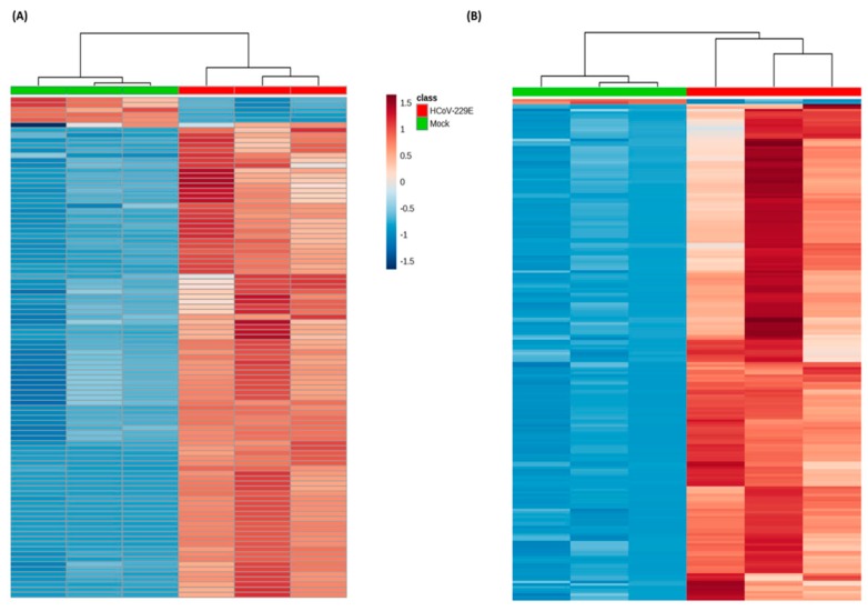
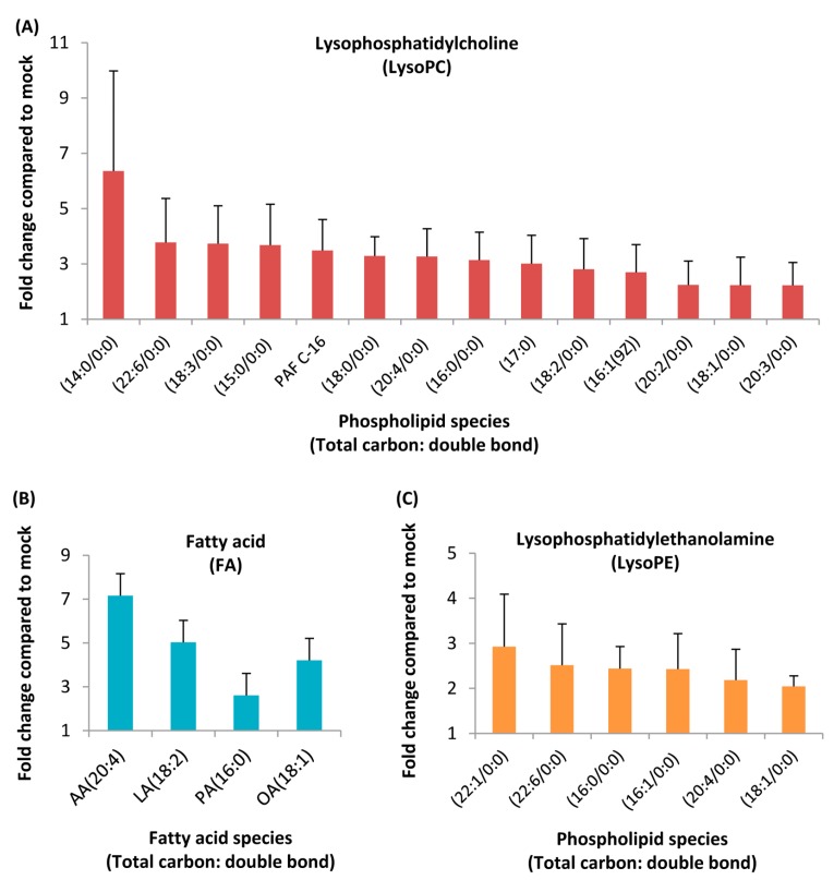
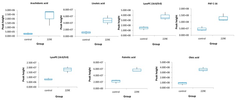
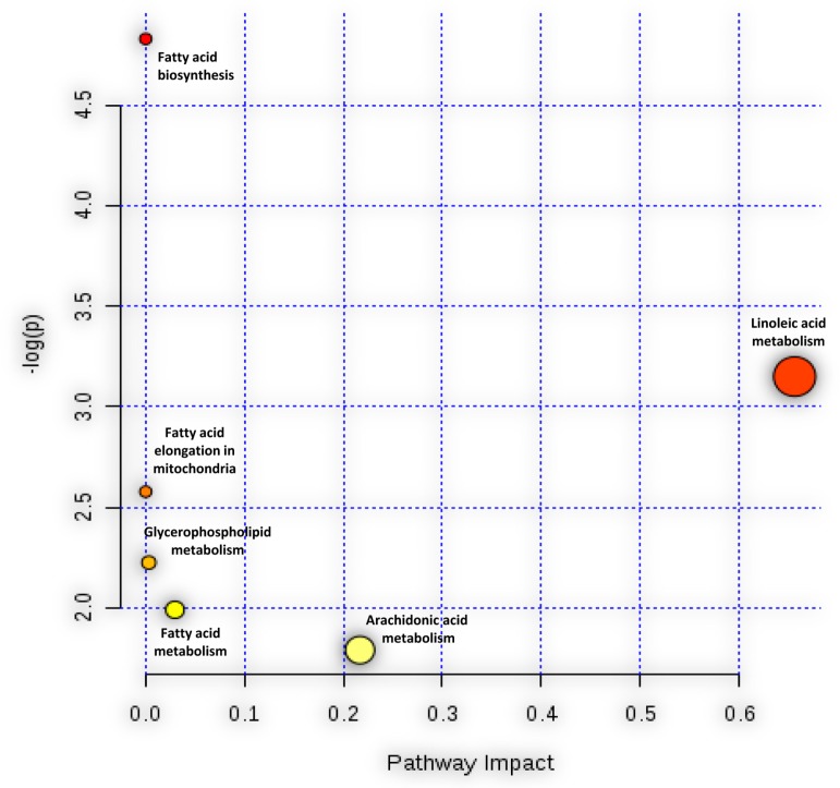
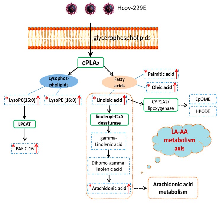
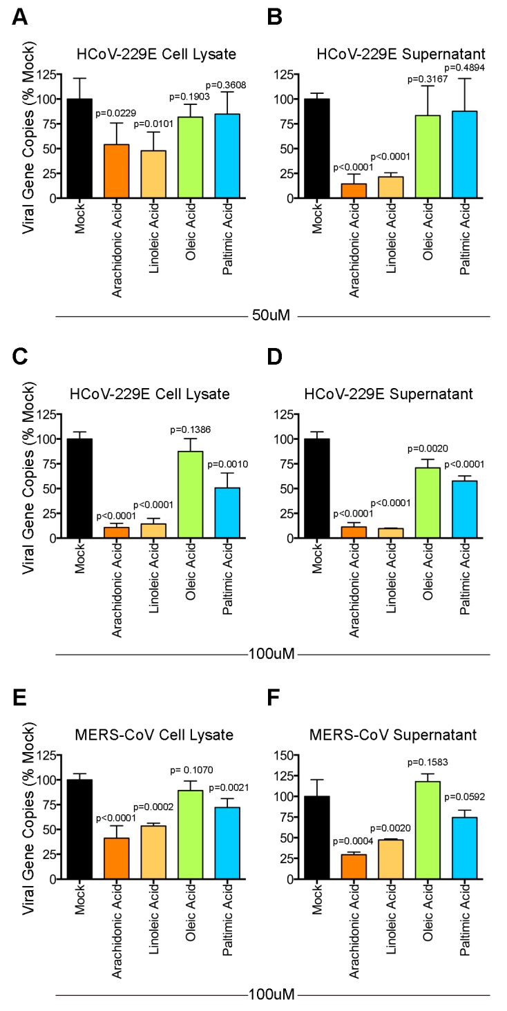
Lipids play numerous indispensable cellular functions and are involved in multiple steps in the replication cycle of viruses. Infections by human-pathogenic coronaviruses result in diverse clinical outcomes, ranging from self-limiting flu-like symptoms to severe pneumonia with extrapulmonary manifestations. Understanding how cellular lipids may modulate the pathogenicity of human-pathogenic coronaviruses remains poor. To this end, we utilized the human coronavirus 229E (HCoV-229E) as a model coronavirus to comprehensively characterize the host cell lipid response upon coronavirus infection with an ultra-high performance liquid chromatography-mass spectrometry (UPLC⁻MS)-based lipidomics approach. Our results revealed that glycerophospholipids and fatty acids (FAs) were significantly elevated in the HCoV-229E-infected cells and the linoleic acid (LA) to arachidonic acid (AA) metabolism axis was markedly perturbed upon HCoV-229E infection. Interestingly, exogenous supplement of LA or AA in HCoV-229E-infected cells significantly suppressed HCoV-229E virus replication. Importantly, the inhibitory effect of LA and AA on virus replication was also conserved for the highly pathogenic Middle East respiratory syndrome coronavirus (MERS-CoV). Taken together, our study demonstrated that host lipid metabolic remodeling was significantly associated with human-pathogenic coronavirus propagation. Our data further suggested that lipid metabolism regulation would be a common and druggable target for coronavirus infections.

Keywords: HCoV-229E; MERS-CoV; UHPLC–MS; lipidomics.


Conflict of interest statement

J.F.-W.C. has received travel grants from Pfizer Corporation Hong Kong and Astellas Pharma Hong Kong Corporation Limited, and was an invited speaker for Gilead Sciences Hong Kong Limited and Luminex Corporation. The other authors declared no conflict of interest. The funding sources had no role in study design, data collection, analysis or interpretation or writing of the report. The corresponding author had full access to all the data in the study and had final responsibility for the decision to submit for publication.

Figures

Figure 1
Figure 1
Heatmap showing the lipidomic analysis of human coronavirus 229E (HCoV-229E)-infected versus non-infected Huh-7 cells. Each rectangle represents an ion feature colored by its normalized intensity scale from blue (decreased level) to red (increased level). The dendrogram on the top was constructed based on the lipid intensity (similarity measure using Euclidean, and the Ward clustering algorithm). HCoV-229E, HCoV-229E-infected cells; Mock, non-infected cells. (A) Significant ion features in negative detection mode; (B) significant ion features in positive detection mode.
Figure 2
Figure 2
Liquid chromatography-mass spectrometry (LC/MS) analysis of HCoV-229E-infected cells revealed a homeostatic change in lipid levels. Huh-7 cells were mock- or HCoV-229E-infected and harvested at 24 hpi. The peak heights of these lipids were calculated and the fold change plotted with GraphPad Prism 5. (A) Lysophosphatidylcholine (LysoPC), (B) fatty acid (FA), (C) lysophosphatidylethanolamine (LysoPE). AA, arachidonic acid; LA, linoleic acid; PA, palmitic acid; OA, oleic acid.
Figure 3
Figure 3
Box-whisker plots of the 7 standard confirmed lipids that were distinguished between the HCoV-229E-infected samples and the non-infected controls. The peak height was generated by LC-MS raw data. Control, non-infected cells; 229E, HCoV-229E-infected cells.
Figure 4
Figure 4
Pathway analysis associate with HCoV-229E infection was carried out by MetaboAnalyst. The Y-axis, “log(p)”, represented the transformation of the original p-value calculated from the enrichment analysis. The X-axis, “Pathway Impact”, represented the value calculated from the pathway topology analysis.
Figure 5
Figure 5
The pathway map based on identified lipids and linoleic acid metabolism recorded in the Kyoto Encyclopedia of Genes and Genomes (KEGG) PATHWAY Database. The star mark “*” indicates the lipids could be matched with commercial standards and have an up-regulation trend. The red arrow represents the up-regulation trend. The blue dashed rectangle and green solid rectangles represent lipids and corresponding enzyme in this pathway, respectively. The orange dashed line represents the LA–AA metabolism axis.
Figure 6
Figure 6
Modulatory effect of lipids on HCoV-229E and Middle East respiratory syndrome coronavirus (MERS-CoV). Huh-7 cells were infected with HCoV-229E. After 1 h of inoculation, the virus inoculum was replaced with medium containing 50 µM (A,B) or 100 μM (C,D) of lipids and incubated for 24 h. The supernatants and cell lysates were collected for reverse-transcription quantitative polymerase chain reaction (RT-qPCR) analysis. In parallel, Huh-7 cells were infected with MERS-CoV. After 1 h of inoculation, the virus inoculum was replaced with medium containing 100 μM (E,F) of lipids and incubated for 24 h. The supernatants and cell lysates were collected for RT-qPCR analysis. Statistical significance was determined by Student’s t-test by comparing the individual lipid-treated group with the mock-treated group (n = 4). The difference was considered significant when p < 0.05.

Similar articles

Cited by

References

    1. Chan J.F., Li K.S., To K.K., Cheng V.C., Chen H., Yuen K.Y. Is the discovery of the novel human betacoronavirus 2c EMC/2012 (HCoV-EMC) the beginning of another SARS-like pandemic? J. Infect. 2012;65:477–489. doi: 10.1016/j.jinf.2012.10.002. - DOI - PMC - PubMed
    1. Chan J.F., To K.K., Tse H., Jin D.Y., Yuen K.Y. Interspecies transmission and emergence of novel viruses: lessons from bats and birds. Trends Microbiol. 2013;21:544–555. doi: 10.1016/j.tim.2013.05.005. - DOI - PMC - PubMed
    1. Chan J.F., Lau S.K., Woo P.C. The emerging novel Middle East respiratory syndrome coronavirus: The “knowns” and “unknowns”. J. Formos Med. Assoc. 2013;112:372–381. doi: 10.1016/j.jfma.2013.05.010. - DOI - PMC - PubMed
    1. Chan J.F., Lau S.K., To K.K., Cheng V.C., Woo P.C., Yuen K.Y. Middle East respiratory syndrome coronavirus: Another zoonotic betacoronavirus causing SARS-like disease. Clin. Microbiol. Rev. 2015;28:465–522. doi: 10.1128/CMR.00102-14. - DOI - PMC - PubMed
    1. Cheng V.C., Lau S.K., Woo P.C., Yuen K.Y. Severe acute respiratory syndrome coronavirus as an agent of emerging and reemerging infection. Clin. Microbiol. Rev. 2007;20:660–694. doi: 10.1128/CMR.00023-07. - DOI - PMC - PubMed

Publication types