

Cervix Stromal Cells and the Progesterone Receptor A Isoform Mediate Effects of Progesterone for Prepartum Remodeling
- PMID: 30654718
- PMCID: PMC6728580
- DOI: 10.1177/1933719118820462
Cervix Stromal Cells and the Progesterone Receptor A Isoform Mediate Effects of Progesterone for Prepartum Remodeling
Abstract
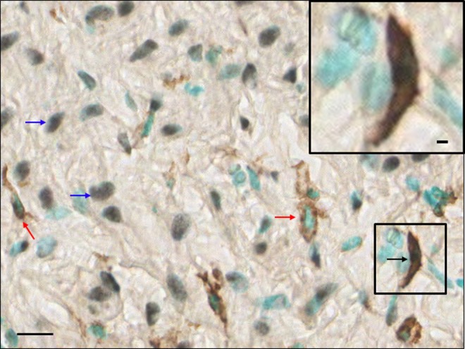
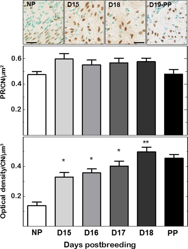
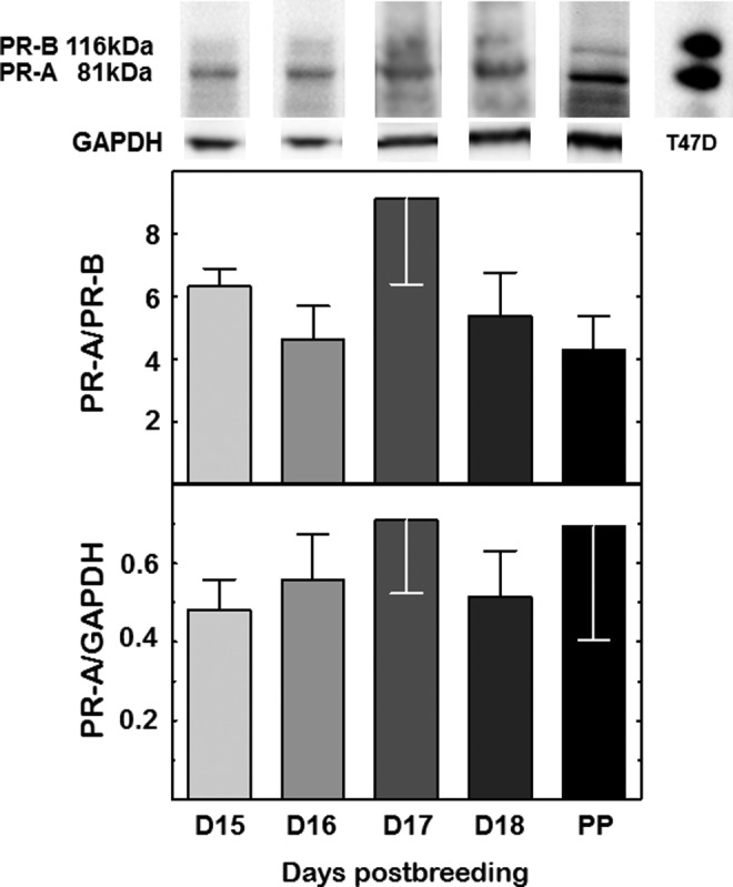
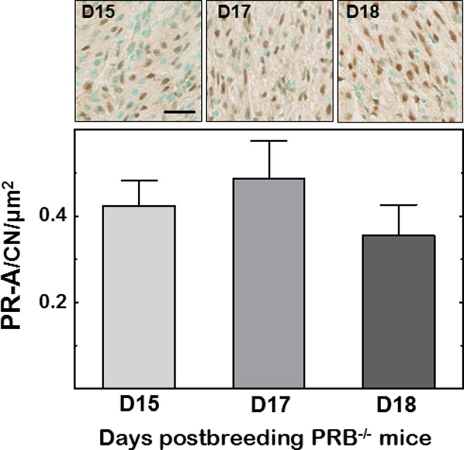
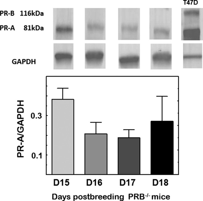
The prepartum transition from a soft to ripening cervix is an inflammatory process that occurs well before birth when systemic progesterone is near peak concentration. This 2-part study first determined that stromal fibroblasts but not macrophages in the cervix have progesterone receptors (PRs). Neither the number of PR cells in cervix sections nor the relative abundance or ratio of nuclear PR isoforms (PR-A/PR-B) were diminished in mice between day 15 of pregnancy and term. Second in mice lacking PR-B ( Pgrtm20mc), the number of cells that expressed the PR-A isoform were maintained during this period of prepartum cervix remodeling. Thus, progesterone effects to sustain pregnancy, as well as soften and ripen the cervix, are mediated by a stable stromal cell population that expresses PR-A and, through interactions with resident macrophages, are likely to mediate inflammatory ripening processes in preparation for birth.
Keywords: mice; cervix; collagen degradation; macrophage; parturition; ripening.
Conflict of interest statement
Figures
Similar articles
-
Progesterone Receptor-Mediated Actions Regulate Remodeling of the Cervix in Preparation for Preterm Parturition.Reprod Sci. 2016 Nov;23(11):1473-1483. doi: 10.1177/1933719116650756. Epub 2016 May 27. Reprod Sci. 2016. PMID: 27233754 Free PMC article.
-
Remodeling of the cervix and parturition in mice lacking the progesterone receptor B isoform.Biol Reprod. 2011 Sep;85(3):498-502. doi: 10.1095/biolreprod.111.091983. Epub 2011 May 25. Biol Reprod. 2011. PMID: 21613631 Free PMC article.
-
Parturition and recruitment of macrophages in cervix of mice lacking the prostaglandin F receptor.Biol Reprod. 2008 Mar;78(3):438-44. doi: 10.1095/biolreprod.107.063404. Epub 2007 Nov 14. Biol Reprod. 2008. PMID: 18003949 Free PMC article.
-
Immunobiology of Cervix Ripening.Front Immunol. 2020 Jan 24;10:3156. doi: 10.3389/fimmu.2019.03156. eCollection 2019. Front Immunol. 2020. PMID: 32038651 Free PMC article. Review.
-
Dynamics of cervical remodeling during pregnancy and parturition: mechanisms and current concepts.Semin Reprod Med. 2007 Jan;25(1):69-79. doi: 10.1055/s-2006-956777. Semin Reprod Med. 2007. PMID: 17205425 Review.
Cited by
-
Recruitment of monocytes and mature macrophages in mouse pubic symphysis relaxation during pregnancy and postpartum recovery†.Biol Reprod. 2019 Aug 1;101(2):466-477. doi: 10.1093/biolre/ioz107. Biol Reprod. 2019. PMID: 31201427 Free PMC article.
-
Contributions to the dynamics of cervix remodeling prior to term and preterm birth.Biol Reprod. 2017 Jan 1;96(1):13-23. doi: 10.1095/biolreprod.116.142844. Biol Reprod. 2017. PMID: 28395330 Free PMC article. Review.
-
Spontaneous preterm birth: Involvement of multiple feto-maternal tissues and organ systems, differing mechanisms, and pathways.Front Endocrinol (Lausanne). 2022 Oct 13;13:1015622. doi: 10.3389/fendo.2022.1015622. eCollection 2022. Front Endocrinol (Lausanne). 2022. PMID: 36313741 Free PMC article. Review.
-
The selective progesterone receptor modulator-promegestone-delays term parturition and prevents systemic inflammation-mediated preterm birth in mice.Am J Obstet Gynecol. 2022 Feb;226(2):249.e1-249.e21. doi: 10.1016/j.ajog.2021.08.013. Epub 2021 Aug 19. Am J Obstet Gynecol. 2022. PMID: 34418351 Free PMC article.
-
Promegestone Prevents Lipopolysaccharide-Induced Cervical Remodeling in Pregnant Mice.Cells. 2025 Feb 7;14(4):242. doi: 10.3390/cells14040242. Cells. 2025. PMID: 39996716 Free PMC article.
References
-
- Word RA, Li XH, Hnat M, Carrick K. Dynamics of cervical remodeling during pregnancy and parturition: mechanisms and current concepts. Semin Reprod Med. 2007;25(1):69–79. - PubMed
-
- Merlino AA, Welsh TN, Tan H, et al. Nuclear progesterone receptors in the human pregnancy myometrium: evidence that parturition involves functional progesterone withdrawal mediated by increased expression of progesterone receptor-A. J Clin Endocrinol Metab. 2007;92(5):1927–1933. - PubMed
Publication types
MeSH terms
Substances
Grants and funding
LinkOut - more resources
Full Text Sources
Research Materials
