. 2019 Jan 17;19(1):27.
doi: 10.1186/s12862-019-1353-z.

Origin and diversification of the plasminogen activation system among chordates

Affiliations

Origin and diversification of the plasminogen activation system among chordates

Andrés Chana-Muñoz et al. BMC Evol Biol. .

Abstract

Background: The plasminogen (PLG) activation system is composed by a series of serine proteases, inhibitors and several binding proteins, which together control the temporal and spatial generation of the active serine protease plasmin. As this proteolytic system plays a central role in human physiology and pathophysiology it has been extensively studied in mammals. The serine proteases of this system are believed to originate from an ancestral gene by gene duplications followed by domain gains and deletions. However, the identification of ancestral forms in primitive chordates supporting these theories remains elusive. In addition, evolutionary studies of the non-proteolytic members of this system are scarce.

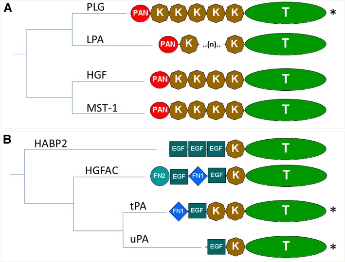
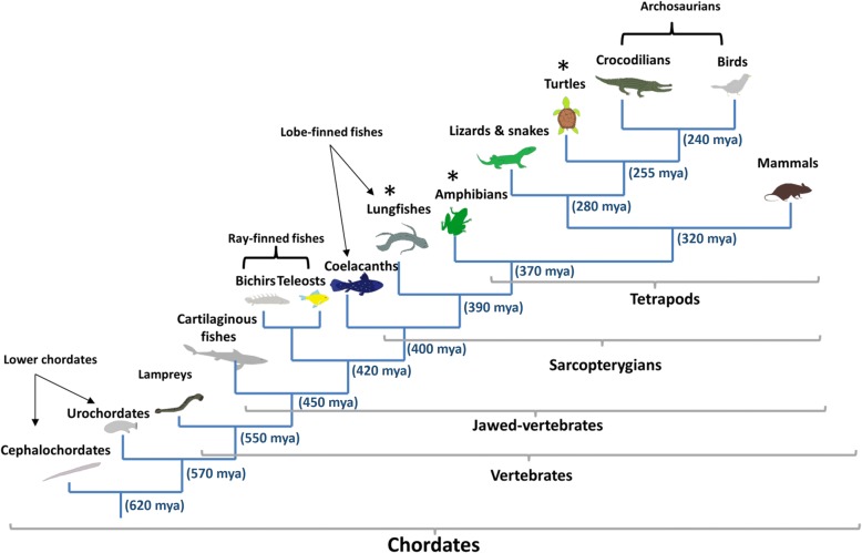
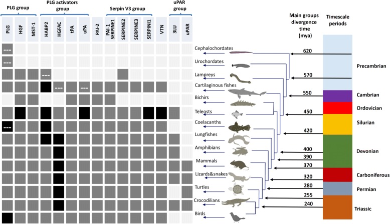
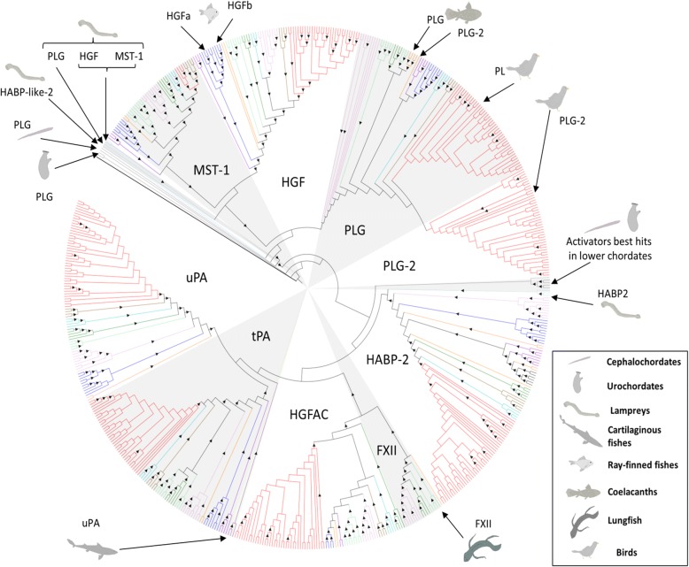
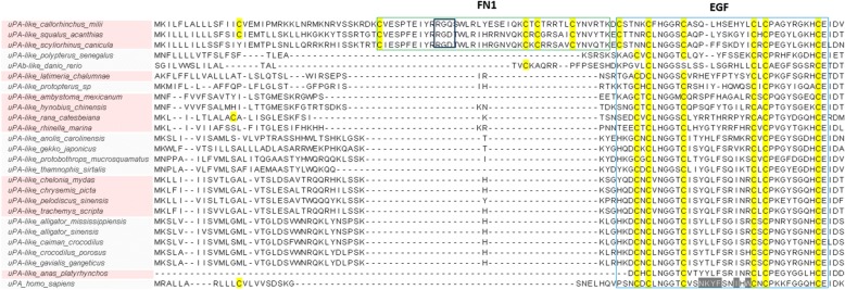
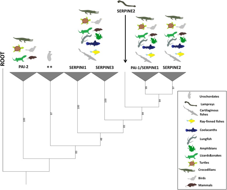
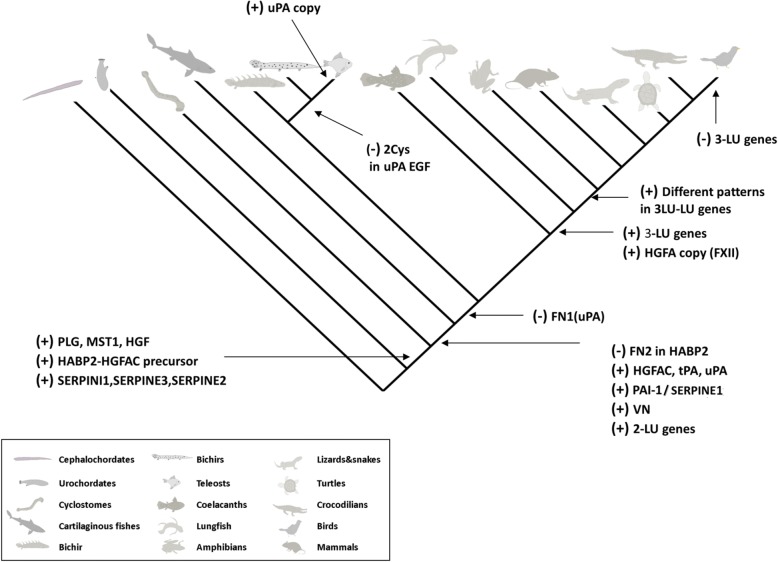
Results: Our phylogenetic analyses place lamprey PLG at the root of the vertebrate PLG-group, while lamprey PLG-related growth factors represent the ancestral forms of the jawed-vertebrate orthologues. Furthermore, we find that the earliest putative orthologue of the PLG activator group is the hyaluronan binding protein 2 (HABP2) gene found in lampreys. The prime plasminogen activators (tissue- and urokinase-type plasminogen activator, tPA and uPA) first occur in cartilaginous fish and phylogenetic analyses confirm that all orthologues identified compose monophyletic groups to their mammalian counterparts. Cartilaginous fishes exhibit the most ancient vitronectin of all vertebrates, while plasminogen activator inhibitor 1 (PAI-1) appears for the first time in cartilaginous fishes and is conserved in the rest of jawed vertebrate clades. PAI-2 appears for the first time in the common ancestor of reptiles and mammals, and represents the latest appearing plasminogen activator inhibitor. Finally, we noted that the urokinase-type plasminogen activator receptor (uPAR)-and three-LU domain containing genes in general-occurred later in evolution and was first detectable after coelacanths.

Conclusions: This study identifies several primitive orthologues of the mammalian plasminogen activation system. These ancestral forms provide clues to the origin and diversification of this enzyme system. Further, the discovery of several members-hitherto unknown in mammals-provide new perspectives on the evolution of this important enzyme system.

Keywords: Chordates; Evolution; Phylogenetic analysis; Plasminogen; Plasminogen activation system; Transcriptome analysis.


Conflict of interest statement

Ethics approval

The animals were euthanized according to the guidelines provided by the Danish Animal Experiments Inspectorate (DAEI). Euthanasia for scientific purposes, such as the harvest of tissue for in vitro studies, did not require permits from the DAEI.

Consent for publication

All authors approved the final manuscript.

Competing interests

The authors declare that they have no competing interests.

Publisher’s Note

Springer Nature remains neutral with regard to jurisdictional claims in published maps and institutional affiliations.

Figures

Fig. 1
Fig. 1
Phylogenetic relationship and protein domain composition of the human serine protease members of the plasminogen activation system and their close paralogues. a PLG-group and b PLG activators group members. PLG (plasminogen), HGF (hepatocyte growth factor), MST-1 (macrophage stimulating 1), HABP2 (hyaluronan binding protein 2), LPA (lipoprotein a), HGFAC (hepatocyte growth factor activator), tPA (tissue-type plasminogen activator) and uPA (urokinase-type plasminogen activator). (*) Members of PAS in mammals. Domain composition: FN1 (fibronectin type 1), FN2 (fibronectin type 2), K (kringle), EGF (epidermal growth factor), PAN (PAN/APPLE), T (trypsin). Phylogenetic tree derived from the human phylome from PhylomeDB [36]
Fig. 2
Fig. 2
Phylogeny of the chordate main groups. Vertebrate groups as described in [54] from which new data was generated (*). Approximate divergence dates are expressed in million years ago (mya) and were collected from [–59]
Fig. 3
Fig. 3
Presence of plasminogen activation system members and related paralogues among the chordate species investigated. Squares indicate absence (light grey), presence (dark grey) and duplication (black) of the different orthologues. Dashed-lines (---) indicate non-canonical protein domain composition. Approximate divergence time in million years ago (mya) was collected from [–59]. PLG (plasminogen), HGF (hepatocyte growth factor), MST-1 (macrophage stimulating 1), HABP2 (hyaluronan binding protein 2), HGFAC (hepatocyte growth factor activator), tPA (tissue-type plasminogen activator) and uPA (urokinase-type plasminogen activator), VTN (vitronectin), uPAR (urokinase-typle plasminogen activator receptor), LU (Ly6/LU domain)
Fig. 4
Fig. 4
Maximum-likelihood phylogenetic tree of the PLG and PLG activator groups of serine proteases. Caenorhabditis elegans SVH-1 (BAL45941.1) was chosen as outgroup to root the tree. Triangles within branches represent bootstrap support higher than 50% after 1000 replicates. Colored branches indicate lower chordates (grey), cartilaginous fishes (violet), ray-finned fishes (blue), coelacanths and lungfish (orange), amphibians (light green), lizards and snakes (dark green), turtles (turquoise), crocodilians (brown) and birds (red). Position of the non-canonical or first appeared candidates is indicated within the tree by arrows. PLG-2 corresponds to the extra PLG gene identified, which has lost the catalytic activity. PLG (plasminogen), HGF (hepatocyte growth factor), MST-1 (macrophage stimulating 1), HABP2 (hyaluronan binding protein 2), HGFAC (hepatocyte growth factor activator), tPA (tissue-type plasminogen activator), uPA (urokinase-type plasminogen activator) and FXII (coagulation factor XII/Hageman factor). The unrooted tree with bootstrap values is provided in Additional file 3
Fig. 5
Fig. 5
Multiple sequence alignment of the N-terminal region of selected vertebrate uPAs. uPA (urokinase-type plasminogen activator), FN1 (fibronectin type 1), EGF (epidermal growth factor)
Fig. 6
Fig. 6
Phylogenetic relationship serpin V3 members and PAI-2. Maximum-likelihood tree rooted at SPN-1 Nematostella vectensis (XP_001627732.1). Percentage of bootstrap support after 1000 replicates shown in the branches. Asterisks (**) indicate the location of reciprocal best hits to human PAI-1 and SERPINI1 in urochordates. PAI-1/SERPINE1 (plasminogen activator inhibitor-1/serpin E1), SERPINE3 (serpin E3), SERPINI1 (serpin I1), SERPINE3 (serpin E3), PAI-2 (plasminogen activator inhibitor-2, placental type). The unrooted tree is provided in Additional file 4
Fig. 7
Fig. 7
Phylogenetic tree of the different proteins containing three LU domains identified in vertebrates and close paralogues. Percentage of bootstrap support after 1000 replicates is shown in each branch. The tree was rooted at the elasmobranch 2 LU branch for convenience in display. a uPAR BRBH in elasmobranchs, b Elephant shark (cartilaginous fish) uPAR BRBH, c BRBH to ray-finned fishes uPAR and PINLYP, coelacanth uPAR and lungfish PINLYP. d Branch containing all the sarcopterygian three-LU genes. Numbers displaying the cysteine pattern of their first, second and third LU domains. e Branch containing human uPAR. uPAR (urokinase-type plasminogen activator receptor), LU (Ly6/uPAR domain), PINLYP (phospholipase A2 inhibitor and LY6/PLAUR domain containing protein), CD177 (cluster of differentiation 177)
Fig. 8
Fig. 8
Overview of the appearance and diversification of the plasminogen activation system among chordates. PLG (plasminogen), HGF (hepatocyte growth factor), MST-1 (macrophage stimulating 1), HABP2 (hyaluronan binding protein 2), LPA (lipoprotein a), HGFAC (hepatocyte growth factor activator), tPA (tissue-type plasminogen activator), uPA (urokinase-type plasminogen activator), VN (vitronectin), FN2 (fibronectin type 2), LU (Ly6/uPAR domain), PAI-1/SERPINE1 (plasminogen activator inhibitor-1/serpin E1), SERPINE2 (serpin E2), SERPINI1 (serpin I1), SERPINE3 (serpin E3), FXII (coagulation factor XII/Hageman factor), Cys (cysteine residues)

Similar articles

Cited by

References

    1. Castellino FJ, Ploplis VA. Structure and function of the plasminogen/plasmin system. Thromb Haemost. 2005;93:647–654. doi: 10.1160/TH04-12-0842. - DOI - PubMed
    1. Andreasen PA, Egelund R, Petersen HH. The plasminogen activation system in tumor growth, invasion, and metastasis. Cell Mol Life Sci. 2000;57:25–40. doi: 10.1007/s000180050497. - DOI - PMC - PubMed
    1. Andreasen PA, Kjoller L, Christensen L, Duffy MJ. The urokinase-type plasminogen activator system in cancer metastasis: a review. Int J Cancer. 1997;72:1–22. doi: 10.1002/(SICI)1097-0215(19970703)72:1<1::AID-IJC1>3.0.CO;2-Z. - DOI - PubMed
    1. Connolly BM, Choi EY, Gardsvoll H, Bey AL, Currie BM, Chavakis T, et al. Selective abrogation of the uPA-uPAR interaction in vivo reveals a novel role in suppression of fibrin-associated inflammation. Blood. 2010;116:1593–1603. doi: 10.1182/blood-2010-03-276642. - DOI - PMC - PubMed
    1. Ploug M, Ronne E, Behrendt N, Jensen AL, Blasi F, Dano K. Cellular receptor for urokinase plasminogen activator. Carboxyl-terminal processing and membrane anchoring by glycosyl-phosphatidylinositol. J Biol Chem. 1991;266:1926–1933. - PubMed

Publication types