

Unusual manifestation of disseminated herpes simplex virus type 2 infection associated with pharyngotonsilitis, esophagitis, and hemophagocytic lymphohisitocytosis without genital involvement
- PMID: 30654754
- PMCID: PMC6337778
- DOI: 10.1186/s12879-019-3721-0
Unusual manifestation of disseminated herpes simplex virus type 2 infection associated with pharyngotonsilitis, esophagitis, and hemophagocytic lymphohisitocytosis without genital involvement
Abstract
Background: Herpes simplex virus (HSV) has various presentations, depending on the patient's immune status, age, and the route of transmission. In adults, HSV type 1 is found predominantly in the oral area, and HSV type 2 (HSV-2) is commonly found in the genital area. HSV-2 infection without genital lesions is uncommon. Herein we report a unique case of pharyngotonsillitis as an initial manifestation of disseminated HSV-2 infection without genital involvement.
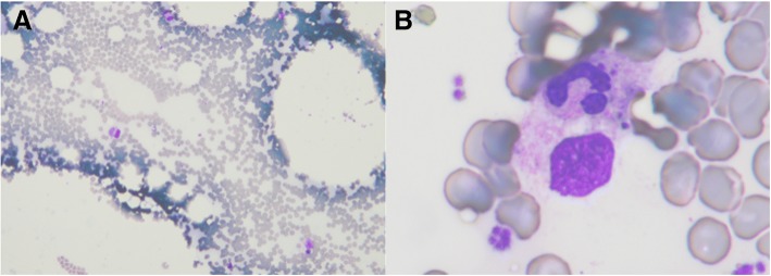
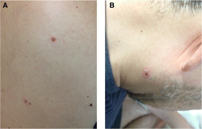
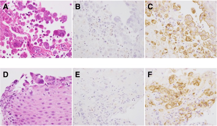
Case presentation: A 46-year-old male was admitted to our hospital with a 1-week history of fever and sore throat. His past medical history included hypereosinophilic syndrome diagnosed at age 45 years. Physical examination revealed throat congestion, bilaterally enlarged tonsils with exudates, tender cervical lymphadenopathy in the left posterior triangle, and mild epigastric tenderness. The laboratory data demonstrated bicytopenia, elevated liver enzyme levels, and hyperferritinemia. A bone marrow smear showed hypocellular marrow with histiocytes and hemophagocytosis. The diagnosis of HLH was confirmed, and the patient was treated with methylprednisolone pulse therapy on days 1-3. On day 5, despite initial improvement of the fever and sore throat, multiple, new, small bullae developed on the patient's face, trunk, and extremities. Additional testing showed that he was positive for HSV-specific immunoglobulin M and immunoglobulin G. Disseminated HSV infection was suspected, and intravenous acyclovir (10 mg/kg every 8 h) was begun. A subsequent direct antigen test of a bulla sample was positive for HSV-2. Moreover, tonsillar and esophageal biopsies revealed viral inclusion bodies. Immunohistochemical staining and a quantitative real-time polymerase chain reaction (PCR) assay confirmed the presence of HSV-2. Disseminated HSV-2 infection with multiple bullae, tonsillitis, esophagitis, and suspected hepatic involvement was diagnosed. After a 2-week course of intravenous acyclovir, his hematological status and liver function normalized, and his cutaneous skin lesions resolved. He was discharged on day 22 in good general health and continued taking oral valacyclovir for viral suppression due to his immunosuppressed status.
Conclusion: Disseminated HSV-2 infection should be considered as one of the differential diagnoses in patients with pharyngotonsillitis and impaired liver function of unknown etiology even if there are no genital lesions.
Keywords: Disseminated infection; Hemophagocytic lymphohistiocytosis; Herpes simplex virus type 2.
Conflict of interest statement
Ethics approval and consent to participate
Not applicable.
Consent for publication
Written informed consent for publication was obtained from the patient.
Competing interests
The authors declare that they have no competing interest.
Publisher’s Note
Springer Nature remains neutral with regard to jurisdictional claims in published maps and institutional affiliations.
Figures
References
Publication types
MeSH terms
Substances
LinkOut - more resources
Full Text Sources
Medical
Miscellaneous
